


CRT is an essential treatment for patients with heart failure and reduced ejection fraction as it can restore left ventricular (LV) electrical and mechanical synchrony. It has been shown to increase quality of life, improve functional status, reduce hospitalisation, improve LV systolic function and reduce mortality in properly selected patients.1,2 While CRT is an effective therapy, approximately 30% of patients treated with CRT do not benefit from it and some patients are negative responders. Improving outcomes with CRT begins with appropriate selection.
QRS Duration and Morphology
Since CRT targets electrical dyssynchrony, QRS duration and morphology have been used to determine which patients will receive maximum benefit from CRT. Based on subgroup analysis of large CRT trials, current guidelines consider CRT implants to be a Class I indication in patients with left bundle branch block (LBBB) and QRS >150 msec, with softer recommendations for QRS <150 msec and non-LBBB patients.3
An analysis of data from the Multicenter Automatic Defibrillator Implantation Trial-Cardiac Resynchronization Therapy (MADIT-CRT) trial demonstrated that only patients with LBBB had a reduction in heart failure events and that non-LBBB patients may have been harmed by CRT.4 Similar data were published from the Resynchronization/Defibrillation for Ambulatory heart Failure Trial (RAFT).5 No benefits were found to result from CRT in right bundle branch block (RBBB) patients in five randomised controlled clinical trials or in a subset of 1,233 patients with non-LBBB QRS morphology from four randomised trials.6,7 However, data on QRS morphology are mixed and no large CRT trial has used QRS morphology as an enrolment criterion. There is no standardised definition of LBBB, especially with respect to predicting electrical dyssynchrony and response to CRT. In addition, some patients with RBBB and intraventricular conduction delay may have a LBBB-like activation pattern of the left ventricle and could respond to CRT.8 To this end, the MADIT-CRT trial showed that, in patients with non-LBBB morphologies and PR intervals >230 msec, there was a 67% reduction in risk of the combined primary endpoint of heart failure and death and a 76% reduction in the risk of death in the CRT device (CRT-D) arm versus the ICD-only arm.9 Data from the REsynchronization reVErses Remodeling in Systolic left vEntricular dysfunction (REVERSE) trial also showed benefit in patients with RBBB receiving CRT.10
QRS duration is a good predictor of CRT response. QRS duration is a criterion for every large clinical trial showing benefit in CRT. Trials randomising patients with narrow QRS (<120 msec or 130 msec) show no benefit or potential harm for patients receiving CRT, even in the presence of mechanical dyssynchrony.11 An analysis of four trials by Cleland et al. showed that QRS duration was the best predicator of benefit from CRT placement, irrespective of QRS morphology, with response seen once QRS duration was >130 msec.12 Subgroup analyses have shown that the greatest CRT benefit is derived in the cohort of patients with a QRS duration of 150 msec, as reflected in the guidelines for CRT placement.3,10,12
Since correcting dyssynchrony is the core benefit of CRT, imaging has been added to 12-lead ECG to expand and refine the population of patients that might benefit from this therapy. The presence of mechanical dyssynchrony on echocardiography or MRI has been shown to predict which patients perform best after CRT placement.13,14 However, the Predictors of Response to CRT (PROSPECT) trial, which prospectively studied echocardiographic measures of dyssynchrony, found they had only modest sensitivity and specificity to predict CRT response with significant variability in the measurement of dyssychrony studied.15 It was conjectured that this was due to the complexity of the dyssynchrony parameters and significant interobserver variability. Newer measures of mechanical dyssynchrony, as assessed by echocardiography and MRI, have shown promise.13,14
Apical rocking and septal flash are simple visual echocardiographic parameters of a LBBB-like contraction pattern that is characterised by late contraction of the lateral left ventricle. Apical rocking specifically refers to a short septal motion of the apex due to early contraction of the septum and late contraction of the lateral wall.13 Septal flash is defined by early contraction of the septum causing a short rapid inward motion of the septum.13
The Relationship of Visually Assessed Apical Rocking and Septal Flash to Response and Long-term Survival Following CRT (PREDICT-CRT) trial assessed 1,060 patients for these parameters and found a 15% reduction in LV end-systolic volume in 77% of patients when both apical rocking and septal flash were present in contrast to 69% if only apical rocking was present and 56% in those with septal flash alone. These parameters were more predictive of echocardiographic response to CRT and long-term survival than QRS morphology, duration and other clinical variables.13 Shoal et al. used cardiac MRI to assess the usefulness of the U-shaped contraction pattern, defined by a line of block in mechanical contraction between the septum and lateral left ventricle, consistent with a true LBBB contraction pattern, to predict CRT outcome. Patients who had a U-shaped propagation had an 80% response rate versus a 26% response rate in the group with homogenous propagation group (p<0.001).16 Similarly, patients without dyssynchrony on MRI, based on a circumferential uniformity ratio estimate >0.70, had no clinical benefit with CRT and a 12-fold increased mortality rate.14
Invasive mapping studies have shown that QRS duration and morphology may not always be predictive of prolonged LV activation times that correlate with CRT response. Non-invasive ECG mapping techniques have been used to identify patients who may have abnormally late-activating regions of the left ventricle. In the Markers and Response to CRT (MARC) study of 240 patients who were followed prospectively, vectorcardiography-derived QRSarea was shown to be more predictive of echocardiographic response to CRT than QRS morphology and duration. The vectorcardiography was mathematically constructed from a standard digital 12-lead ECG and consists of three orthogonal leads X, Y, and Z that form a 3D vector loop.17
In addition, high-resolution non-invasive electrocardiographic imaging (ECGI) has shown promise in defining patients who are more likely to respond to CRT. Multiple studies have shown that ECGI measures of electrical dyssynchrony obtained using the CardioInsight™ system (Medtronic) better correlate with acute haemodynamic and long-term clinical response to CRT than the presence of LBBB.18 Another smaller ECGI system with a 53-electrode ECG belt (Heartscape Technologies) has also shown the ability to predict echocardiographic response to CRT better than QRS duration or morphology.19
Lead Placement
Another strategy for improving CRT outcome is optimisation of lead placement. Since the lateral left ventricle is the latest-activating area in LBBB, the lateral or posterolateral left ventricle – in general – is the preferred target for LV lead placement, but the optimal place for such placement may vary for a given patient. Early data showed that the placement of anterolateral or posterolateral leads was superior to anterior lead placement.20 However, analysis of the Comparison of Medical Therapy, Pacing and Defibrillation in Heart Failure (COMPANION) trial showed that no particular lead location was associated with an improved response.21
Other studies show that apical LV lead placement is associated with worse outcomes than non-apical leads.22 The Targeted Left Ventricular Lead Placement to Guide CRT (TARGET) and Speckle Tracking Assisted Resynchronization Therapy for Electrode Region (STARTER) studies used speckle tracking echocardiography to determine the area of latest mechanical activation for placement of the LV lead. Patients randomised to the targeted LV lead placement group had lower rates of the combined endpoint of heart failure and death.23,24 This approach is limited by the availability of specialised software and the need for optimal image quality.
LV leads should also be targeted to areas of viable myocardium. CRT patients with scar tissue in the posterolateral left ventricle assessed by cardiac MRI have minimal response to CRT.25 Further data consistently show that LV leads placed in areas of viable myocardium – especially in viable segments with dyssynchrony – have high response rates to CRT versus LV leads placed in an area of scar and no dyssynchrony.14
Late-activating segments of the left ventricle can be targeted electrically. LV pacing from areas of late activation defined by either LV lead electrical delay (QLV is defined as the time between onset of the QRS on the surface ECG and the sensed signal on the LV lead) or interventricular delay (time of onset of large positive or negative peaks of the right ventricular to LV electrogram) have been shown to correlate with favourable acute haemodynamic and clinical responses.26,27 Even patients with a lead placed in the LV apex had a good response to CRT if the lead showed late LV activation.28 Leads placed in an electrically late-activating segment predicted by ECGI or vectorcardiography have also been shown to enhance acute response to CRT.28–30 However, preliminary results from the CRT Implant Strategy Using the Longest Electrical Delay for Non-left Bundle Branch Block Patients (ENHANCE–CRT) pilot study, which randomised 248 patients to LV lead placement guided by QLV versus standard LV lead placement in non-LBBB subjects, showed no statistical difference.31
Applicability of CRT therapy can also be limited by the anatomic constraints of the coronary sinus (CS). In 5–10% of patients, LV lead placement is unsuccessful due to either CS inaccessibility, high LV pacing thresholds, or phrenic nerve stimulation.1,2,6 In addition, >50% of patients have only one CS branch that is suitable for lead placement, making it difficult to target LV lead placement to areas of late activation or dyssynchrony in all subjects.32
New Catheter Approaches to LV Synchronisation
Endocardial LV pacing offers many potential benefits over epicardial LV pacing. LV endocardial leads can be targeted to any area of the left ventricle due to the lack of anatomic constraints from the CS. In addition, LV endocardial pacing thresholds are lower than epicardial leads and phrenic nerve stimulation can be more readily avoided. Animal and human studies have shown that the LV endocardium provides a favourable acute haemodynamic response to LV pacing. This is likely to due to more rapid LV endocardial impulse conduction leading to shorter LV activation times as compared with epicardial pacing.33
The most common technique for achieving LV endocardial pacing involves transseptal access across the intra-atrial septum to deliver a LV lead across the mitral annulus into the left ventricle. The largest trial of LV endocardial pacing with transseptal atrial placement, the ALternate Site Cardiac ResYNChronization (ALSYNC) trial, enrolled 138 patients and had an 89.4% procedural success rate in patients who were CRT non-responders or who had failed implants. The clinical response rate was 59%. However, despite anticoagulation there was a high rate of thromboembolic complications (stroke rate was 2.6 per 100 patient years and there were 14 transient ischaemic attacks in nine patients), but no cases of lead-related mitral regurgitation were seen.34
Another technique for LV endocardial pacing involves passing the lead through the intraventricular septum and into the lateral LV myocardium. In a preliminary study, all patients were successfully implanted with this technique and eight out of nine were considered responders. All patients were on anticoagulation and no cerebrovascular accidents or transient ischaemic attacks were reported during a mean follow-up of 8.7 months.35 The larger Pilot Study of Interventricular Septal Puncture for Cardiac Resynchronization Therapy to Treat Heart Failure (LV-CONSEPT) trial (NCT01818765), designed to evaluate outcomes for this approach to LV lead placement, has completed enrolment.
Preliminary data from LV endocardial pacing via the LV septum has also been shown to improve haemodynamics versus right ventricular pacing and to have similar haemodynamic response to lateral LV endocardial biventricular pacing. This technique utilises a specialised pacing lead with a fixed 4 mm helix. The helix has a thin coating on the proximal portion, with only the distal 1.27 mm electrode exposed. The lead is screwed into the intraventricular septum until LV septal capture is confirmed. The advantage of this technique is that the lack of hardware in the left ventricle eliminates the need for anticoagulation.36
The final option for LV endocardial pacing is leadless pacing. The WiSE™ CRT System (EBR Systems) uses a leadless 9 mm pacing electrode directly implanted into the LV endocardium via retrograde or transseptal access. The electrode is powered by a generator implanted near the left ventricle in the intercostal space that delivers ultrasound to the electrode, which is then converted to electricity for pacing. The LV electrode will endothelialise, so long-term anticoagulation is not required. In the preliminary Safety and Performance of Electrodes Implanted in the Left Ventricle (SELECT-LV) study of 35 patients, there was a 97% success rate for implantation, with an 85% clinical response rate to CRT in patients that failed CRT implants or were non-responders. However, 23% of participants in this trial experienced significant adverse events.37 The larger Simulation Of the Left Ventricular Endocardium for CRT (SOLVE-CRT) trial (NCT02922036) will look at 350 patients who are non-responders or who have failed CRT implants to assess the utility of this system.
Another option to improve CRT response is to pace the LV from two different sites. A study looking at 40 patients with permanent AF with a slow ventricular response randomised patients to conventional CRT versus CRT with two LV leads and showed higher LV ejection fraction (27% versus 35%) and smaller LV end-systolic volume (157 cm3 versus 134 cm3) with dual site LV pacing.38 A second study compared 34 patients who had received CRT with two LV leads to a propensity score-matched population that had received conventional CRT.39 The conventional CRT group had more frequent ventricular tachycardia and a higher all-cause mortality and heart transplant rate. Preliminary data from the 100-patient Triple-site versus Standard Cardiac Resynchronization Therapy (TRUST-CRT) study showed similar rates of adverse events and implantation success with triple-site versus standard CRT and an improved rate of clinical response, with only 12.5% of patients reporting New York Heart Association (NYHA) Class III symptoms versus 30% in the standard CRT group. However, triple-site CRT was associated with higher LV lead thresholds, lower pacing impedance and greater battery drain.40 No further data have been published, although the trial completed enrolment in 2015. The recently published the triple-site CRT(V3) study showed that in 84 CRT non-responders randomised to a second LV lead versus control, there was no improvement in clinical response to CRT in the treatment group at 24 months, but there was a high rate (20%) of procedural complications.41 Multiple randomised controlled trials of dual-site LV pacing have been completed but have not yet released their results.
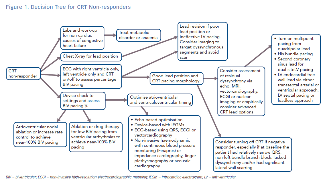
Multisite LV pacing can also be delivered via a quadripolar LV lead. Multiple point pacing (MPP) is an option in most commercially available CRT systems and allows for dual-site LV pacing by using two separate bipoles from a quadripolar lead. By covering a larger area of LV myocardium, MPP can increase the speed of impulse propagation and reduce LV activation times. MPP has shown improvement over single-site LV pacing as assessed by pressure volume loops, LV dP/dtmax, global peak LV radial strain and LV outflow tract velocity time integral in selected patients.42 In the MultiPoint Pacing IDE (MPP-IDE) study, MPP delivered from anatomically-separate bipoles resulted in an 87% clinical response rate and 100% response rate in patients who were non-responders at 3 months. MPP may be a good option to use in patients that do not initially respond to standard CRT therapy.43 Figure 1 outlines a strategy for improving the clinical response of CRT non-responders.
His bundle pacing is emerging as a first-line option for optimal CRT response in non-responders and in those with unfavourable CS anatomy. It is based on the premise that longitudinal dissociation exists in the proximal His bundle and disease within this bundle causes bundle branch block. Stimulation of the distal His bundle can narrow and potentially normalise a widened QRS. Animal and human studies from >40 years ago showed that temporary pacing of the distal His bundle resulted in QRS normalisation.44,45 More recent work has shown that it is feasible to permanently pace the His with standard pacing leads and that His bundle pacing can result in narrowing of the QRS in 70–90% of patients.46–48 In initial work, Lustagarten et al. noted similar outcomes between His bundle pacing and CS lead pacing for CRT, with significantly shorter procedure times with the former therapy.49 A larger series reported by Sharma et al. showed a 90% success rate in narrowing the QRS with His bundle pacing.50 The clinical response rate was 70% and mean ejection fraction increased from 30% to 44%. There was also a significant improvement in NYHA class, with participants’ symptoms improving by one NYHA class on average. Another recent series of 39 patients with RBBB from the same group showed that His bundle pacing was successful in 95% of patients and there was a favourable clinical response in 76% of patients.50 His bundle pacing offers many potential advantages:
- Faster impulse propagation due to endocardial and His–Purkinje system recruitment;46,47
- Direct access to the anatomic area of interest;46
- Lack of need for optimisation of CRT lead placement or ventriculoventricular (VV) timing, since recruitment of the distal His-Purkinje system and fascicles should result in normalisation of LV electrical activation;49,50
- Endocardial pacing with QRS narrowing should avoid the negative CRT response that is sometimes associated with LV epicardial pacing due to ventricular proarrhythmia or unchanged dyssynchrony pattern.51
While His bundle pacing offers promise as a first-line therapy, current drawbacks include:
- lack of randomised trials showing clinical improvement or mortality benefit;
- high lead revision rates;
- higher thresholds and current battery drain associated than for CRT; and
- lack of significant long-term data of lead performance in the His position.
The on-going His Bundle Pacing Versus Coronary Sinus Pacing for CRT (His-SYNC) pilot trial (NCT02700425) will randomise 40 patients to CRT with His bundle pacing versus a LV lead. Further large clinical trials are required before this form of pacing can be considered a first-line method for CRT.
Narrow QRS
Narrow QRS patients are generally excluded from CRT due to the lack of benefit demonstrated in multiple large randomised controlled trials. However, selected patients with narrow QRS do benefit from CRT. Many studies have shown the CRT pacing coupled with atrioventricular (AV) node ablation, either with a LV lead or His bundle pacing, have reported improved heart failure symptoms and LV ejection fraction in patients with AF, LV dysfunction and congestive heart failure.52,53 The recently published Abate and Place in AF plus CRT (APAF-CRT) trial randomised 109 patients with a narrow QRS and permanent AF to AV node ablation and CRT versus rate control. The CRT–AV node ablation group had a significant reduction in death from any cause or hospitalisation for heart failure (12% versus 33%) and a trend towards decreased mortality (4% versus 12%).54
While CRT for narrow QRS patients in sinus rhythm is currently contraindicated, some studies have suggested that in selected patients the majority of acute haemodynamic improvement from CRT is due to AV optimisation, thus improving LV preload rather than correcting dyssynchrony.55 The His Optimized Pacing Evaluated for Heart Failure (HOPE-HF) trial (NCT02671903) will randomise 160 patients with a LV ejection fraction <40% with narrow QRS or RBBB and PR interval >200 msec to His bundle pacing with AV optimisation versus standard congestive heart failure therapy, testing the hypothesis that AV optimisation improves outcome in this group of patients.56
Atrioventricular and Ventriculoventricular Timing
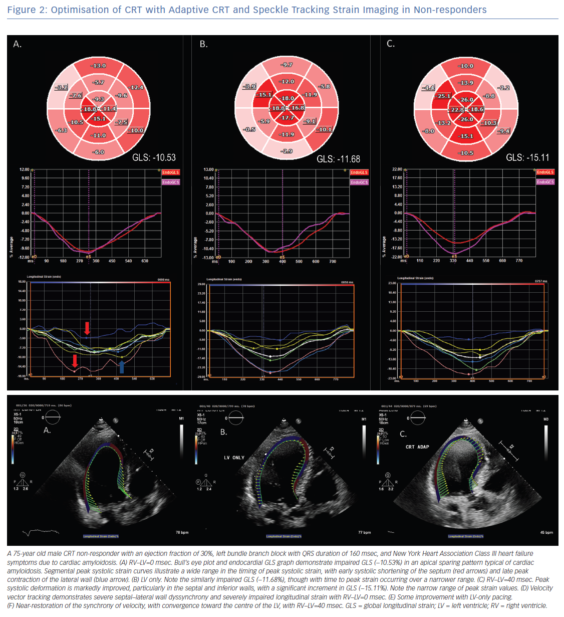
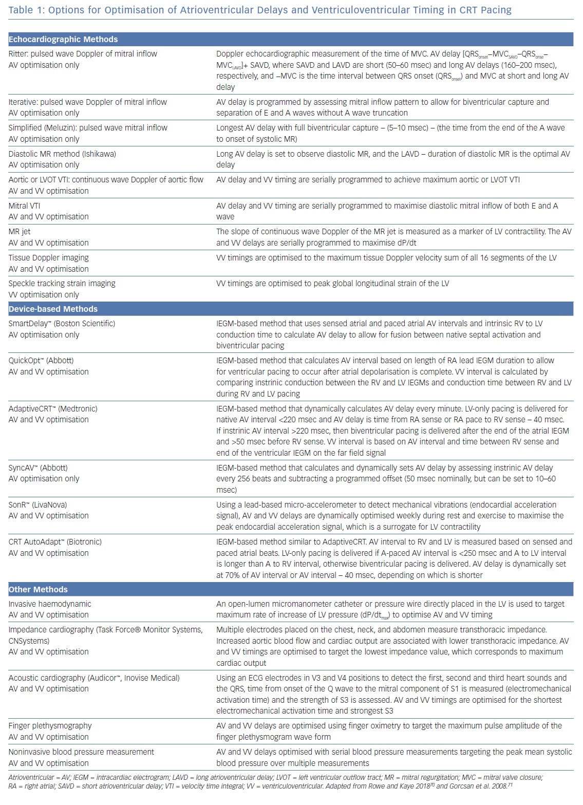
Two initial large trials evaluated the use of AV and VV timing optimisation to improve CRT outcomes – SmartDelay determined AV Optimization: A Comparison of AV Optimization Methods Used in CRT (SMART-AV) and Frequent Optimization Study Using the QuickOpt Method (FREEDOM), but these showed no additional benefit over nominal settings.57 As a consequence, guidelines currently only recommend AV and VV timing optimisation for CRT non-responders. Dynamic algorithms that change programmed settings based on frequent automatic assessments, thereby optimising AV and VV timing, have performed better. Adaptive CRT (aCRT) uses intrinsic conduction to determine timings and results in LV-only pacing with a sensed AV delay <220 msec and biventricular pacing otherwise. The aCRT algorithm was associated with a reduced 30-day readmission rate of 14.8% versus 24.8% in controls. Figure 2 shows optimisation of CRT with echocardiographic strain imaging using aCRT. Patients in the aCRT group with >50% LV-only pacing had an 82% clinical response rate versus 68% in the optimised biventricular pacing group.58
The Clinical Trial of the SonRtip Lead and Automatic AV–VV Optimization Algorithm in the PARADYM RF SonR CRT-D (RESPOND-CRT) trial assessed the clinical utility of the SonR™ (LinaNova) contractility sensor in CRT optimisation. The SonR sensor measures mechanical vibrations, which correlate with LV dP/dTmax. The study randomised 998 patients in a 2:1 fashion to receive weekly automatic CRT optimisation with SonR versus echocardiographic optimisation.59 There was a 75% response rate in the SonR group versus 70% in the control group, with a 35% relative reduction in the risk of hospitalisation for heart failure. The SyncAV™ algorithm (Abbott) regularly calculates the PR interval from device electrograms and automatically adjusts the AV delay to allow for intrinsic septal activation before biventricular pacing. This algorithm shortens QRS duration during biventricular pacing to a greater extent than statically optimised AV and VV delays and LV-only pacing.60 A recent randomised study showed that programming AV delays and VV timing to promote fusion with intrinsic septal activation and targeting the shortest QRS duration with CRT pacing resulted in a higher rate of reverse remodelling than nominal settings (74% versus 53%).61 Table 1 summarises the various methods of AV and VV timing optimisation.
CRT with an ICD
There has been significant debate as to whether the addition of ICD therapy to CRT improves outcomes. The majority of patients who meet the CRT criteria are also suitable for an ICD for the primary prevention of sudden cardiac death. Furthermore, most patients enrolled in the pivotal trials demonstrating CRT benefit also received an ICD.62
Multiple retrospective studies have shown better survival in patients who received a CRT-D rather than a CRT pacemaker (CRT-P).63,64 Additionally, in patients with ischaemic cardiomyopathy, CRT-D implantation was associated with improved mortality over CRT-P.63,64 This may be because the patients who received CRT-P were generally older, more likely female and more often had non-ischaemic cardiomyopathy as well as a greater number of comorbidities, which likely contributed to their lower survival.63,64 In its favour, CRT-P is costs less and has been associated with fewer complications than CRT-D.65 It should be noted that large cohort studies have shown that in patients with non-ischaemic cardiomyopathy, the addition of ICD therapy to CRT is not associated with improved outcomes;66 however, in non-ischaemic cardiomyopathy with scarring, specifically LV mid-wall fibrosis seen with cardiac MRI, CRT-D improves mortality and reduces major adverse cardiac events.67
The Prospective Observational Study of the ICD in Sudden Cardiac Death Prevention (PROSe-ICD) study developed a clinical decision tool using biomarkers and clinical variables to help guide those who would benefit from ICD therapy in addition to CRT.68 The CeRtiTuDe study showed that while patients receiving CRT-P had a higher mortality, 95% of this excess mortality was not due to sudden cardiac death.69 The guidelines currently recommend CRT-P for NYHA Class IV heart failure and favour CRT-D for younger patients with milder heart failure, with ischaemic heart disease and life expectancy >1 year.65 CRT-P is recommended for older patients with more advanced congestive heart failure, severe renal dysfunction, frailty and lower life expectancy.65
Conclusion
CRT is a potent therapy for improving outcomes and reducing mortality in heart failure. Despite being an established first-line therapy, significant issues remain in patient selection and the proper delivery of CRT. On-going research into new tools and methods to improve CRT therapy will allow for further improvements in outcomes in what has already proven an innovative therapy for the treatment of symptomatic LV systolic dysfunction.
Clinical Perspective
- To improve CRT, more accurate patient selection is needed to include new populations that will respond, and limit implantation in patient groups with poor outcomes.
- Better coronary sinus lead placement location and device programming is needed to improve CRT outcomes with existing technology.
- New pacing approaches are required to improve left ventricular synchronisation.