Article

Session 4: Contact Force: What Does It Mean, and How Much Do We Need It?

Register or Login to View PDF Permissions
Permissions× For commercial reprint enquiries please contact Springer Healthcare: ReprintsWarehouse@springernature.com.

For permissions and non-commercial reprint enquiries, please visit Copyright.com to start a request.

For author reprints, please email rob.barclay@radcliffe-group.com.
Information image
Average (ratings)
No ratings
Your rating
Copyright Statement:

The copyright in this work belongs to Radcliffe Medical Media. Only articles clearly marked with the CC BY-NC logo are published with the Creative Commons by Attribution Licence. The CC BY-NC option was not available for Radcliffe journals before 1 January 2019. Articles marked ‘Open Access’ but not marked ‘CC BY-NC’ are made freely accessible at the time of publication but are subject to standard copyright law regarding reproduction and distribution. Permission is required for reuse of this content.

The availability of technologies for real-time contact for sensing has significantly advanced and invigorated techniques of catheter ablation of AF, particularly with RF energy. This session was an in-depth look at contact force and included a critical perspective of the head-tohead with cryoballoon ablation. The experts discussed their opinions on the use of contact for sensing to reduce or avoid lesion gaps, and highlighted the significant and evolving body of evidence of clinical outcomes, as well as the caveats of too much contact force during the AF procedure.

Relevance of Fire and Ice

AF is the most common arrhythmia with a prevalence >33 million patients.20 Approximately 40% are asymptomatic,21 and another 30% are effectively treated with anti-arrhythmia drugs (AAD).22 Of the remaining 30% who are symptomatic and with failed AAD treatment,22 just 4% are treated annually23 – amounting to 396,000 treated with catheter ablation, and leaving 22.7 million patients who have not been effectively treated by drugs or undergone catheter ablation.22,23

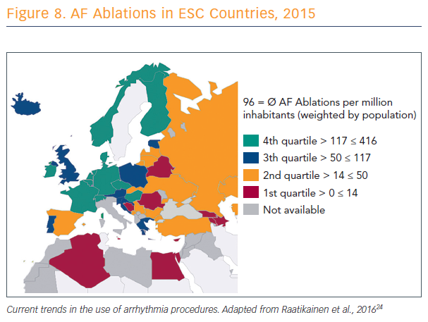
There is a lack of consistency across Europe in numbers and rates of catheter ablation procedures (see Figure 8).24 In these countries, said Prof Karl-Heinz Kuck of Asklepios Klinik St. Georg in Hamburg, Germany, electrophysiologists are discussing what to add to PVI. Countries with lower rates do not have access to catheter ablation or, if they have access, there are no clinicians available who know how to do it.

The development of balloon-based PVI is on the upswing, Prof Kuck suggested, because of the long learning curve, complexity, and challenge to create transmural, contiguous and permanent lesions with point-by-point ablation. He stressed that he is a strong supporter of CF-based catheter ablation, but this technique may not be optimal for every provider, especially given the widespread nature of AF.

AF Ablations in ESC Countries, 2015

Article image

There is no randomised controlled trial showing superiority of catheter ablation with contact force, compared to conventional RF catheter ablation, Prof Kuck explained. The only randomised trial that has compared a control group with a contact force group, aiming at non-inferiority, showed the same success rate when comparing the primary endpoints.25

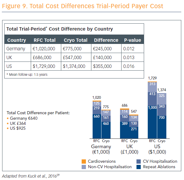
Ablation strategies that target the PVs and/or PV antrum are the cornerstone of most ablation procedures and, if the PVs are targeted, electrical isolation should be the goal. Furthermore, the new ESC guidelines recommend that catheter ablation should target isolation of the pulmonary veins using RF ablation or cryoballoon catheters.9 The radiofrequency catheter uses heat in a focal point-by-point delivery guided by electro-anatomical mapping, and cryoballoon uses freezing in a balloon single-step delivery guided by fluoroscopy without mapping. Prof Kuck explained the parameters for use of both techniques, and discussed the results of the largest cryoballoon versus RF ablation study ever performed and published.26 Cryoballoon ablation resulted in non-inferior mid-term efficacy compared with RF ablation at a median follow-up of 14 months, with significantly lower mean procedure times and lower left atrial (LA) dwell-time and lower costs (see Figure 9). Pericardial effusion was higher in RF patients compared with cryoballoon, but phrenic nerve injury was higher in cryoballoon and there were no incidences of phrenic nerve injury in the RF group. Mean fluoroscopy time was 2 minutes lower in the RF group.

“The cryoballoon-based PVI approach in paroxysmal AF is noninferior to the radiofrequency-based PVI with regard to frequency and safety, and superior with regard to re-hospitalization and re-ablation,” Prof Kuck concluded. “Novel balloon-based radiofrequency ablation systems combine direct visualisation of the anatomical substrate and, thereby, also new technologies for lesion formation. Permanent PVI can potentially be achieved to a high extent with a balloon technology… to my understanding, it’s the future of catheter ablation of AF, and the future is now.”

CF: What Does it Mean and How Does it Affect Lesion?

Traditional parameters of RF lesion control have been the target electrode temperature, delivered radiofrequency power in the power control mode, RF durattimes impedance-based control, and electrodetissue contact – an important but hitherto unquantified parameter. In the previous era, contact was evaluated in various semi-quantitative ways: fluoroscopy, tactile feedback, electrograms, electrode temperature and impedance, and intracardiac echo.

“It is only over the last 7 or 8 years that real-time contact force sensing has been available, which has given precise control of lesion creation,” said Prof Dipen Shah, from University Hospitals Geneva, Switzerland. Electrode tissue contact can be thought of as comprising of two components. The most important is the magnitude of the surface area of the electrode in direct contact with the tissue – the contact footprint.

Atrial tissue and the ventricle and myocardial tissue are soft, and as a rigid electrode is pushed into it, it gets more and more enveloped into the tissue. The second component is the stability of this contact, which can be considered as being composed of spatial stability (sliding of the tip electrode over the endocardium), and temporal stability, maintaining the intensity of the contact over time.

To quantify the contact pressure a catheter tip exerts on the tissue, the operator needs to know the exact surface area of contact. However, this cannot be assessed precisely. Experiments on ex vivo, porcine left atria showed that the same amount of force applied to different parts of the left atrium reduced the wall thickness by different amounts, depending on the thickness and tissue composition of the wall.27

A dynamic situation exists in vivo, including the effects of both cardiac and respiratory movement. To estimate a dynamically changing contact force over time, or at least its intensity, Prof Shah’s team formulated the area under the real-time contact force curve, termed the ‘force-time integral’, as a cumulative index of the amount of force over time. This allows, in one sense, a good measure of temporal stability, whereas spatial stability requires precise, two-dimensional localisation capabilities with respect not to a stable extra-cardiac spatial reference such as a back patch, but with reference to the intracardiac endocardia, which we do not yet have.

Research by Prof Shah and his colleagues determined the general effects of increased CF: increased electrode-tissue interface surface area; reduced electrode surface area exposed to (low impedance) blood; reduced electrode tip-sliding; tissue compression and thinning; tissue trauma; and higher tissue temperatures during RF delivery, including higher probability of a ‘pop’ and higher probability of extracardiac heating.28

To mitigate for risk, contact force sensors should be appropriately zeroed and re-zeroed after every reintroduction into the vascular system or through a sheath. It should be kept in mind that electromagnetic interference is likely to reduce the accuracy of this form of CF sensing. Catheter stability is important, and the composition of the atrial tissue should be kept in mind to reduce variances in lesion size.

Measuring CF remains important for recognising instances of absence of contact, reduction of ineffective ablations, better control of lesion size and repeatability, better arrhythmia-free outcome after PVI for paroxysmal atrial fibrillation (pAF) with optimal contact force, improved specificity for low-voltage substrate, reduced tissue trauma during catheter manipulation, and monitoring influence of respiration on contact, which Prof Shah thinks is an often-underestimated advantage of real-time CF sensing.

Total Cost Differences Trial-Period Payer Cost

Article image

Determinants of Gap in CF-guided PV Encircling

Next, Prof Mattias Duytschaever from Bruges, Belgium, discussed CF–guided point-by-point PV encircling, and what determines a gap when CF has been used according to the ‘rules’. That is, if a patient has a reconnection in one or more veins, what has caused the gap, was there a weak link in the initial ablation chain and, finally, is it clinically relevant?

Prof Duytschaever presented an analysis of research on 42 paroxysmal AF patients who underwent contact force-guided PV encirclement. In the resulting 84 circles, 840 segments were identified, 44 of which had a gap. Segments with a gap were compared to durable segments without a gap.

Prof Duytschaever’s analysis defined the weakest link within each segment as the minimal time of application, power, delta impedance, CF, force-time integral and ablation index, and the maximal inter-lesion distance.

The ablation index is a formula that integrates power and contact force over time, taking into account the rapid rise of the lesion in the first seconds of the ablation, and a greater contribution of power.

“Inter-lesion distance seems straightforward, but don’t forget this is a new tool,” said Prof Duytschaever. “Before, we could not reliably measure inter-lesion distance because we were just putting tags during our ablation.

Prof Mattias Duytschaever presenting

Article image

“The key finding: the difference between a segment with a gap and no gap is a lower ablation index. Vice versa, where all the ablation indexes were to a high level of specificity, the difference between segments with gap and no gap is the inter-lesion distance. They are independent; if you combine both parameters, if inter-lesion distance and the ablation index are more than 400 at the posterior wall and more than 550 on the anterior wall, you have a 93 % specificity for predicting a durable segment.”

Building on these data, Prof Duytschaever’s research group explored the use of enclosing the PVs with ablation index-guided RF applications with an inter-lesion distance of <6 mm and found the reproducible, perfect circle “invariably leading to PV isolation, and that makes a big difference. We [also] consider the presence of the oesophagus – an ablation index of 300 is enough if I have signs of oesophageal injury. That is most of the time an application of around 8 seconds.”

There were two complications in 250 patients using this approach, neither conclusively linked to the procedure, and no significant adverse events.

Prof Duytschaever’s group has submitted a paper on the comparative data of the last 50 CF-guided patients versus the first 50 close-guided ablations with the strict criteria. They have reached near-100 % acute isolation rate.

This is very reproducible; colleagues who are now using this technique, all acknowledge that this is reproducible and superior to CF-guided ablation, he said. Prof Duytschaever’s results show rates of freedom from AF at 3, 6, and 12 months of 92 % in the close-guided group.

Prof Duytschaever concluded that acute and late gaps in contact force–guided PV encircling are due to insufficient lesion depth and/ or discontiguity. The use of the strict-criteria ablation protocol results in a significant improvement in acute durability and arrhythmia-free survival. Nevertheless, real-time assessment of lesion formation remains one of the most important unmet needs in cardiac electrophysiology.

Randomised Trials of CF Efficacy: A Critical Look at the Literature

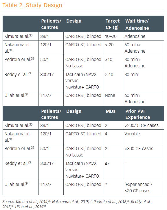
There have been five studies that have randomised patients to having the CF data either available to the operator or blinded to the operator (see Table 2).30–34 CF targets in each of the studies were ‘fairly modest’ at 5–20 g, and all except one involved a long waiting period of 60 minutes to watch for spontaneous reconnection. Most of these studies showed that using contact force shortened ablation time, decreased fluoroscopy time, and resulted in significantly fewer sites of acute reconnection. Complication rates did not differ significantly, but then these were infrequent overall.

Study Design

Article image

“However, what really matters is how these patients did on follow-up, and [the existing randomised studies show] absolutely no improvement in clinical outcomes,” said Dr Dhiraj Gupta of Liverpool Heart and Chest Hospital in the UK.

“That’s really a big disappointment. Why have contact force trials not shown clinical success? Why couldn’t they prove what most operators believe strongly to be true?”

Analysis of the five trials raised two issues:

  • long waiting time (30–60 minutes) – poorer quality lesions created without contact force got a chance to manifest themselves and be re-ablated, thereby negating much of the benefit; and
  • mandating either no CF targets or low CF targets (<20g) – this meant that in most trials, the average CF between the two groups was similar.

“If we’re just concentrating on contact force alone we’re missing the point,” said Dr Gupta. “Contact force data help determine how deep each legion is, but it is equally important to have good contiguity between adjacent lesions. We know that force time integral is superior to contact force as a marker of lesion size. And power is an important component of legion creation. If you use high power, contact force becomes less and less relevant. And that’s where ablation index comes in. Contact force is critical in ablation delivery but, at the end of the day, it’s only one ingredient in what is a very complex recipe.”

References

  1. Zoni-Berisso M, Lercari F, Carazza T, Domenicucci S. Epidemiology of atrial fibrillation: European perspective. Clin Epidemiol 2014;6:213–20.
    Crossref PubMed
  2. Goette A, Kalman JM, Aguinaga L, et al. EHRA/HRS/APHRS/ SOLAECE expert consensus on atrial cardiomyopathies: Definition, characterisation, and clinical implication. J Arrhythm 2016;32:247–78.
    Crossref PubMed
  3. Wijesurendra RS, Liu A, Eichhorn C, et al. Lone atrial fibrillation is associated with impaired left ventricular energetics that persists despite successful catheter ablation. Circulation 2016;134:1068–81.
    Crossref PubMed
  4. Kolb C, Nürnbuger S, Ndrepepa G, et al. Modes of initiation of paroxysmal atrial fibrillation from analysis of spontaneously occurring episodes using a 12-lead Holter monitoring system. Am J Cardiol 2001;88:853–7.
    Crossref PubMed
  5. Ehrlich JR, Cha TJ, Zhang L, et al. Cellular electrophysiology of canine pulmonary vein cardiomyocytes: action potential and ionic current properties. J Physiol 2003;551: 801–13.
    Crossref PubMed
  6. de Groot N, van der Does L, Yaksh A, et al. Direct proof of endo-epicardial asynchrony of the atrial wall during atrial fibrillation in humans. Circ Arrhythm Electrophysiol 2016;9:pii: e003648.
    Crossref PubMed
  7. Kirchof P, Benussi S, Koetecha D, et al. 2016 ESC guidelines for the management of atrial fibrillation developed in collaboration with EACTS. Eur J Cardiothorac Surg 2016;50:e1- e88.
    Crossref PubMed
  8. Pison L, La Meir M, van Opstal A, et al. Hybrid thoracoscopic surgical and transvenous catheter ablation of atrial fibrillation. J Am Coll Cardiol 2012;60:54–61.
    Crossref PubMed
  9. Dudink E, Essers B, Holvoet W, et al. Acute cardioversion vs a wait-and-see approach for recent-onset symptomatic atrial fibrillation in the emergency department: Rationale and design of the randomized ACWAS trial. Am Heart J 2017;183:49–53.
    Crossref PubMed
  10. Ouyang F, Tilz R, Chun J, et al. Long-term results of catheter ablation in paroxysmal atrial fibrillation: Lessons from a 5-year follow-up. Circulation 2010;122:2368–77.
    Crossref PubMed
  11. Calkins H, Reynolds MR, Spector P, et al. Treatment of atrial fibrillation with antiarrhythmic drugs or radiofrequency ablation. Circ Arrythm Electrophysiol 2009;2:349–61.
    Crossref PubMed
  12. de Vos CB, Pisters R, Nieuwlaat R, et al. Progression from paroxysmal to persistent atrial fibrillation: clinical correlates and prognosis. J Am Coll Cardiol 2010;55:725–31.
    Crossref PubMed
  13. Weimar T, Schena S, Bailey MS, et al. The Cox-Maze procedure for lone atrial fibrillation: a single-center experience over 2 decades. Circ Arrhythm Electrophysiol 2012;5:8–14.
    Crossref PubMed
  14. Gallagher MM, Camm AJ. Classification of atrial fibrillation. Pacing Clin Electrophysiol 1997;20:1603–5.
    Crossref PubMed
  15. January CT, Wann LS, Alpert JS. 2014 AHA/ACC/HRS guideline for the management of patients with atrial fibrillation: executive summary: a report of the American College of Cardiology/American Heart Association Task Force on practice guidelines and the Heart Rhythm Society. Circulation 2014;130:2071–104.
    Crossref PubMed
  16. Platonov PG, Mitrofanova LB, Orshanskaya V, et al. Structural abnormalities in atrial walls are associated with presence and persistency of atrial fibrillation but not with age. J Am Coll Cardiol 2011;58:2225–32.
    Crossref PubMed
  17. Kottkamp H, Schreiber D. The substrate in “early persistent” atrial fibrillation: arrhythmia induced, risk factor induced, or from a specific fibrotic atrial cardiomyopathy? J Am Coll Cardiol Clin Electrophysiol 2016;2:140–2.
    Crossref
  18. Kottkamp H, Schreiber D, Moser F, Rieger A. Therapeutic approaches to atrial fibrillation ablation targeting atrial fibrosis. J Am Coll Cardiol EP 2017;3:643–53.
    Crossref
  19. Gianni C, Atoui M, Mohanty S, et al. Difference in thermodynamics between two types of esophageal temperature probes: Insights from an experimental study. Heart Rhythm 2016;13:2195–200.
    Crossref PubMed
  20. Rahman F, Kwan GF, Benjamin EJ. Global epidemiology of atrial fibrillation. Nat Rev Cardiol 2014;11:639–54.
    Crossref PubMed
  21. Steinberg BA, Holmes DN, Ezekowitz MD, et al. Rate versus rhythm control for management of atrial fibrillation in clinical practice: results from the Outcomes Registry for Better Informed Treatment of Atrial Fibrillation (ORBIT-AF) registry. Am Heart J 2013;165:622-9.
    Crossref PubMed
  22. Calkins H, Reynolds MR, Spector P, et al. Treatment of atrial fibrillation with antiarrhythmic drugs or radiofrequency ablation: two systematic literature reviews and metaanalyses. Circ Arrhythm Electrophysiol 2009;2:349-61.
    Crossref PubMed
  23. Medtronic internal estimates
  24. Raatikainen MJ, Arnar DO, Merkely B, et al. Access to and clinical use of cardiac implantable electronic devices and interventional electrophysiological procedures in the European Society of Cardiology Countries: 2016 Report from the European Heart Rhythm Association. Europace 2016;18(Suppl 3):iii1–iii79.
    Crossref PubMed
  25. Reddy VY, Dukkipati SR, Neuzil P, et al. Randomized, controlled trial of the safety and effectiveness of a contact force-sensing irrigated catheter for ablation of paroxysmal atrial fibrillation: Results of the TactiCath Contact Force Ablation Catheter Study for Atrial Fibrillation (TOCCASTAR) Study. Circulation 2015;132:907–15.
    Crossref PubMed
  26. Kuck KH, Brugada J, Fürnkranz A. Cryoballoon or radiofrequency ablation for paroxysmal atrial fibrillation. N Engl J Med 2016;374:2235–45.
    Crossref PubMed
  27. Wright M, Harks E, Kolen A, et al. Contact force is a poor marker of tissue compression in the left atrium. Utility of a novel intra-tissue visualization & ablation system to assess tissue depth in real time. Europace 2014;16(Suppl 2):9–4, ii5
  28. Shah DC, Mandar M. Real-time contact force measurement: a key parameter for controlling lesion creation with radiofrequency energy. Circ Arrhythm Electrophysiol 2015;8:713– 21.
    Crossref PubMed
  29. Chun KRJ, Brugada J, Elvan A, et al. The Impact of Cryoballoon Versus Radiofrequency Ablation for Paroxysmal Atrial Fibrillation on Healthcare Utilization and Costs: An Economic Analysis From the FIRE AND ICE Trial. J Am Heart Assoc 2017;6:pii: e006043.
    Crossref PubMed
  30. Kimura M, Sasaki S, Owada S, et al. Comparison of lesion formation between contact force-guided and non-guided circumferential pulmonary vein isolation: a prospective, randomized study. Heart Rhythm 2014;11:984–91.
    Crossref PubMed
  31. Nakamura K, Naito S, Sasaki T, et al. Randomized comparison of contact force-guided versus conventional circumferential pulmonary vein isolation of atrial fibrillation: prevalence, characteristics, and predictors of electrical reconnections and clinical outcomes. J Interv Card Electrophysiol 2015;44:235– 45.
    Crossref PubMed
  32. Pedrote A, Arana-Rueda E, Arce-León A, et al. Impact of contact force monitoring in acute pulmonary vein isolation using an anatomic approach. A randomized study. Pacing Clin Electrophysiol 2016;39:361–9.
    Crossref PubMed
  33. Reddy VY, Dukkipati SR, Neuzil P, et al. Randomized, controlled trial of the safety and effectiveness of a contact force-sensing irrigated catheter for ablation of paroxysmal atrial fibrillation: results of the TactiCath Contact Force Ablation Catheter Study for Atrial Fibrillation (TOCCASTAR) Study. Circulation 2015;132:907–15.
    Crossref PubMed
  34. Ullah W, McLean A, Tayebjee MH, et al. Randomized trial comparing pulmonary vein isolation using the SmartTouch catheter with or without real-time contact force data. Heart Rhythm 2016;13:1761–7.
    Crossref PubMed
  35. Perna F, Heist EK, Danik SB, et al. Assessment of catheter tip contact force resulting in cardiac perforation in swine atria using force sensing technology. Circ Arrhythm Electrophysiol 2011;4:218–24.
    Crossref PubMed
  36. Quallich SG, Van Heel M, Iaizzo PA. Optimal contact forces to minimize cardiac perforations before, during, and/or after radiofrequency or cryothermal ablations. Heart Rhythm 2015;12:291–6.
    Crossref PubMed
  37. Yokoyama K, Kakagawa H, Shah DC, et al. Novel contact force sensor incorporated in irrigated radiofrequency ablation catheter predicts lesion size and incidence of steam pop and thrombus. Circ Arrhythm Electrophysiol 2008;1:354–62.
    Crossref PubMed
  38. Kuck KH, Reddy VY, Schmidt B, et al. A novel radiofrequency ablation catheter using contact force sensing: Toccata study. Heart Rhythm 2012;9:18–23.
    Crossref PubMed
  39. Natale A, Reddy VY, Monir G, et al. Paroxysmal AF catheter ablation with a contact force sensing catheter: results of the prospective, multicenter SMART-AF trial. J Am Coll Cardiol 2014;64:647–56.
    Crossref PubMed
  40. Perino A, Fan J, Schmitt S, et al. Cost variation and associated outcomes of catheter ablation for atrial fibrillation. J Am Coll Cardiol 2015;65(10S):A277" target="_blank">PubMed
  41. Ho SY, Sanchez-Quintana D, Cabrera JA, Anderson RH. Anatomy of the left atrium: implications for radiofrequency ablation of atrial fibrillation. J Cardiovasc Electrophysiol 1999;10:1525–33.
    CrossrefPubMed
  42. Nakamura K, Funabashi N, Uehara M, et al. Left atrial wall thickness in paroxysmal atrial fibrillation by multislice-CT is initial marker of structural remodeling and predictor of transition from paroxysmal to chronic form. Int J Cardiol 2011;148:139–47.
    Crossref PubMed
  43. Platonov PG, Ivanov V, Ho SY, Mitrofanova L. Left atrial wall thickness in patients with and without atrial fibrillation. J Cardiovasc Electrophysiol 2008;9:689–92.
    Crossref PubMed
  44. Pan NH, Tsao HM, Chang NC, et al. Aging dilates atrium and pulmonary veins. Chest 2008;133:190–6.
    Crossref PubMed
  45. Whitaker J, Rajani R, Chubb H, et al. The role of myocardial wall thickness in atrial arrhythmogenesis. Europace 2016;18:1758–72.
    Crossref PubMed
  46. Mukherjee RK, Chubb H, Harrison JL, et al. Epicardial electroanatomical mapping and radiofrequency ablation in the swine left ventricle under real time MRI guidance. Heart Rhythm 2017;14(Suppl):S191
  47. Jumrussirikul P, Atiga WL, Lardo AC, et al. Prospective comparison of lesions created using a multipolar microcatheter ablation system with those created using a pullback approach with standard radiofrequency ablation in the canine atrium. Pacing Clin Electrophysiol 2000;23:203–13.
    Crossref PubMed
  48. Avitall B, Helms RW, Koblish JB, et al. The creation of linear contiguous lesions in the atria with an expandable loop catheter. J Am Coll Cardiol 1999;33:972–84.
    Crossref PubMed
  49. van Rensburg H, Willems R, Holemans P, et al. Simultaneous creation and evaluation of linear radiofrequency lesions. J Interv Card Electrophysiol 2002;6: 215–24.
    PubMed
  50. Gepstein L, Hayam G, Shpun S, et al. Atrial linear ablations in pigs. Circulation 1999;100:419–26.
    Crossref PubMed
  51. Schwartzman D, Michele JJ, Trankiem CT, Ren JF. Electrogramguided radiofrequency catheter ablation of atrial tissue comparison with thermometry-guide ablation: comparison with thermometry-guide ablation. J Interventional Cardiac Electrophysiol 2001;5:253–66.
    PubMed
  52. Bortone A, Brault-Noble G, Appetiti A, Marijon E. Elimination of the negative component of the unipolar atrial electrogram as an in vivo marker of transmural lesion creation: acute study in canines. Circ Arrhythm Electrophysiol 2015;8:905–11.
    Crossref PubMed
  53. Zghaib T, Ipek EG, Zahid S, et al. Association of left atrial epicardial adipose tissue with electrogram bipolar voltage and fractionation: Electrophysiologic substrates for atrial fibrillation. Heart Rhythm 2016;13:2333–9.
    Crossref PubMed
  54. Iwasaki YK, Nishida K, Kato T, Nattel S. Atrial fibrillation pathophysiology: implications for management. Circulation 2011;124:2264–74.
    Crossref PubMed
  55. Khurram IM, Habibi M, Gucuk IE, et al. Left atrial LGE and arrhythmia recurrence following pulmonary vein isolation for paroxysmal and persistent AF. JACC Cardiovasc Imaging 2016;9:142–8.
    Crossref
  56. Habibi M, Lima JA, Gucuk IE, et al. The association of baseline left atrial structure and function measured with cardiac magnetic resonance and pulmonary vein isolation outcome in patients with drug-refractory atrial fibrillation. Heart Rhythm 2016;13:1037–44.
    Crossref PubMed
  57. Di Biase L, Burkhardt D, Mohanty P, et al. Periprocedural stroke and management of major bleeding complications in patients undergoing catheter ablation of atrial fibrillation: The impact of periprocedural therapeutic international normalized ratio. Circulation 2010;121:2550–6.
    Crossref PubMed
  58. Di Biase L, Burkhardt JD, Santangeli P, et al. Periprocedural stroke and bleeding complications in patients undergoing catheter ablation of atrial fibrillation with different anticoagulation management. Circulation 2014;129:2638–44.
    Crossref PubMed
  59. Calkins H, Willems S, Gerstenfeld EP, et al. Uninterrupted dabigatran versus warfarin for ablation in atrial fibrillation. N Engl J Med 2017;376:1627–36.
    Crossref PubMed