Article

Clinical Evidence Demonstrating the Benefit of MultiPoint Pacing Cardiac Resynchronisation Therapy

Register or Login to View PDF Permissions
Permissions× For commercial reprint enquiries please contact Springer Healthcare: ReprintsWarehouse@springernature.com.

For permissions and non-commercial reprint enquiries, please visit Copyright.com to start a request.

For author reprints, please email rob.barclay@radcliffe-group.com.
Information image
Average (ratings)
No ratings
Your rating

Abstract

Cardiac resynchronisation therapy (CRT) using biventricular pacing is an established therapy for impairment of left ventricular (LV) systolic function in patients with heart failure (HF). Although technological advances have improved outcomes in patients undergoing biventricular pacing, the optimal placement of pacing leads remains challenging, and approximately one third of patients have no response to CRT. This may be due to patient selection and lead placement. Electrical mapping can greatly improve outcomes in CRT and increase the number of patients who derive benefit from the procedure. MultiPoint™ pacing (St Jude Medical, St Paul, MN, US) using a quadripolar lead increases the possibility of finding the best pacing site. In clinical studies, use of MultiPoint pacing in HF patients undergoing CRT has been associated with haemodynamic and clinical benefits compared with conventional biventricular pacing, and these benefits have been sustained at 12 months. This article describes the proceedings of a satellite symposium held at the European Heart Rhythm Association (EHRA) Europace conference held in Milan, Italy, in June 2015.

Disclosure:Dr Curnis has received fees from Medtronic Inc, Biotronik,St Jude Medical, Boston Scientific, Sorin Liva Nova and Spectranetics; Dr O’Donnell has received fellowship support from Medtronic Inc and St Jude Medical, advisory booard fees from St Jude Medical and Metronic and speakers fees from Medtronic Inc, St Jude Medical, Boston Scientific, Boehringer Ingelheim and Merit Medical; Dr Kloppe has received consulting fees from Medtronic Inc and St Jude Medical, and lecture fees from Medtronic Inc, Boston Scientific and St. Jude Medical; Dr Calovic has received speakers fees from St Jude Medical

Received:

Accepted:

Published online:

Support:This supplement is based on the proceedings of a St Jude Medical satellite symposium held at the EHRA Europace conference, Milan, Italy in June 2015. This report was written by a medical writer, Katrina Mountfort, and the participants all reviewed and approved this supplement before publication. St Jude Medical reviewed the supplement for technical accuracy and sponsored the development of this supplement in Arrhythmia & Electrophysiology Review.

Copyright Statement:

The copyright in this work belongs to Radcliffe Medical Media. Only articles clearly marked with the CC BY-NC logo are published with the Creative Commons by Attribution Licence. The CC BY-NC option was not available for Radcliffe journals before 1 January 2019. Articles marked ‘Open Access’ but not marked ‘CC BY-NC’ are made freely accessible at the time of publication but are subject to standard copyright law regarding reproduction and distribution. Permission is required for reuse of this content.

Dr Kloppe began his presentation by discussing a 2008 multicentre single-blind crossover study that randomised patients with congestive HF (n=40) either to triventricular (one RV and two LV leads) or biventricular (one RV and one LV lead) stimulation. The primary endpoint was quality of ventricular resynchronisation (Z ratio). The study did not find a significant change in Z ratio but concluded that CRT with triple-site ventricular stimulation was safe and resulted in a significantly higher LVEF and smaller LV end-systolic volume and diameter.24

Since this study, quadripolar leads have been developed. A 2013 study (n=21) compared biventricular and multisite pacing with a quadripolar LV lead. Using an acute pacing protocol, four configurations of multisite and simultaneous RV pacing were tested. In the majority (84 %) of patients, MultiPoint pacing improved LV dP/dt(max) compared with biventricular pacing.25 Only three patients with MultiPoint pacing (one who was a super-responder) experienced no significant additional effect from MultiPoint pacing.

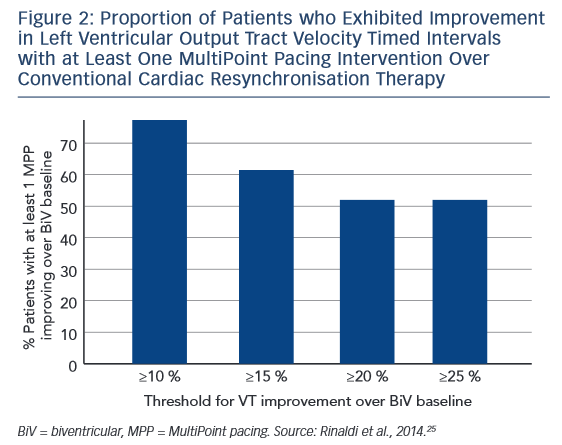
In a prospective multicentre study (n=40), a CRT defibrillator incorporating a quadripolar lead was programmed to deliver MultiPoint pacing with eight different configurations of timing delays. The configuration that yielded the best echocardiographic measurement for each patient was defined as ‘optimal MultiPoint pacing’. The endpoint was analysis of contractility expressed as global radial strain of the opposing walls and stroke volume LV output tract velocity timed intervals (LVOT VTIs). Compared with conventional CRT, the mean peak radial strain was significantly higher for the optimal MultiPoint pacing configuration (18.3 ± 7.4 versus 9.3 ± 5.3 %; p<0.001).26 The median delay between LV1 and LV2 was 55 ms, and between LV2 and RV was 20 ms. In a subanalysis, no difference was seen between ischaemic and nonischaemic patients. The proportion of patients who exhibited improvements in LVOT VTI with at least one MultiPoint pacing intervention over conventional CRT is shown in Figure 2, and suggests that low and non-responders might derive more benefit from MultiPoint pacing.

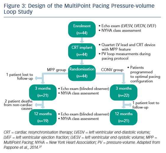
Most recently, a study by Pappone et al. investigated MultiPoint pacing in 44 consecutive patients in an acute setting. Following CRT implantation, consecutive patients (n=44) were randomised to receive biventricular pacing with either conventional LV pacing (CONV) or MultiPoint pacing (see Figure 3). In each case, an optimal pacing configuration was determined based on intra-operative pressure-volume (PV) loop measurements. The primary endpoint was reduction in LV end-systolic volume (LVESV) of ≥15 %. The delay between LV1 and LV2 was 26 ± 15 ms and between LV2 and RV was 10 ± 9 ms. After 3 months, 50 % of CONV patients and 76 % of MultiPoint pacing patients were classified as responders. ESV reduction, EF increase and NYHA class reduction relative to baseline were significantly greater in the MultiPoint pacing group than in the CONV group. In terms of aetiology, non-ischaemic patients had a significant reduction in LVESV compared with ischaemic patients, but no differences were seen in other parameters. A significant increase in MultiPoint pacing group in contractility, stroke volume and EF was seen with MultiPoint pacing. In addition, the best MultiPoint pacing improved acute diastolic function, i.e., change from baseline.27 In 2015, Pappone published 12-month data from this study, showing sustained improvements; these are discussed in the final presentation.28

Proportion of Patients who Exhibited Improvement in Left Ventricular Output Tract Velocity Timed Intervals with at Least One MultiPoint Pacing Intervention Over Conventional Cardiac Resynchronisation Therapy

Article image

Design of the MultiPoint Pacing Pressure-volume Loop Study

Article image

Another recent study compared biventricular pacing and MultiPoint pacing in a group of 29 patients with a high QRS. The effect of MultiPoint pacing, by means of simultaneous pacing from distal and proximal dipoles, was investigated at any available site, an average of 3.2 pacing sites per patients, and a total of 92 measurements. An increase in LV electrical delay (Q-LV) at any site was seen even with MultiPoint pacing relative to biventricular pacing when taking the best and worst sites. Q-LV was measured at each location, along with the increase in LVdP/ dtmax (maximum rate of rise of LV pressure) obtained by biventricular pacing and MultiPoint pacing. The effect of MultiPoint pacing, by means of simultaneous pacing from distal and proximal dipoles, was investigated at all available sites. Compared with biventricular pacing at any LV site, MultiPoint pacing yielded a small but consistent increase in haemodynamic response. A correlation between the increase in haemodynamics and Q-LV on MultiPoint pacing was observed for all measurements, including those taken at the best and worst sites. The MultiPoint pacing-induced improvement in contractility was associated with significantly greater narrowing of the QRS complex than conventional biventricular pacing. A good correlation was observed between electrical delay and haemodynamic improvement.29

In an ongoing study (n=19), Kloppe et al. measured the acute effect of conventional pacing (SSP) and MultiPoint pacing on dP/dt(max) compared with baseline. A significant increase was observed with MultiPoint pacing relative to SSP but wide inter-patient response to the two interventions was observed. At the recent Heart Rhythm Society meeting, preliminary data were presented from a multicentre prospective study (n=313) that received MultiPoint pacing or biventricular pacing. In the MultiPoint pacing group, EF increased significantly compared with biventricular (42±8 % versus 35 ± 10 %; p<0.001).30

In conclusion, a growing body of clinical data have demonstrated that CRT with MultiPoint pacing can significantly improve acute cardiac contractility and haemodynamic parameters compared with conventional pacing. MultiPoint pacing can further augment the well-described systolic benefits of CRT, likely by better synchronising LV contraction and/or recruiting additional LV myocardial tissue. The MultiPoint pacing-induced improvement in contractility was associated with significantly greater narrowing of the QRS complex than conventional biventricular pacing, However, much remains poorly understood, including the optimal timing of LV1 to LV2 to RV.

References

  1. Epstein AE, DiMarco JP, Ellenbogen KA, et al. 2012 ACCF/AHA/ HRS focused update incorporated into the ACCF/AHA/HRS 2008 guidelines for device-based therapy of cardiac rhythm abnormalities: a report of the American College of Cardiology Foundation/American Heart Association Task Force on Practice Guidelines and the Heart Rhythm Society. J Am Coll Cardiol 2013;61:e6–75.
  2. Bristow MR, Saxon LA, Boehmer J, et al. Cardiacresynchronization therapy with or without an implantable defibrillator in advanced chronic heart failure. N Engl J Med 2004;350:2140–50.
  3. Cleland JG, Daubert JC, Erdmann E, et al. The effect of cardiac resynchronization on morbidity and mortality in heart failure. N Engl J Med 2005;352:1539–49.
  4. Moss AJ, Hall WJ, Cannom DS, et al. Cardiac-resynchronization therapy for the prevention of heart-failure events. N Engl J Med 2009;361:1329–38.
  5. Curtis AB, Yancy CW, Albert NM, et al. Cardiac resynchronization therapy utilization for heart failure: findings from IMPROVE HF. Am Heart J 2009;158:956–64.
  6. Perrin MJ, Green MS, Redpath CJ, et al. Greater response to cardiac resynchronization therapy in patients with true complete left bundle branch block: a PREDICT substudy. Europace 2012;14:690–5.
  7. Zareba W, Klein H, Cygankiewicz I, et al. Effectiveness of Cardiac Resynchronization Therapy by QRS Morphology in the Multicenter Automatic Defibrillator Implantation Trial- Cardiac Resynchronization Therapy (MADIT-CRT). Circulation 2011;123:1061–72.
  8. O‘Donnell D, Lin T, Swale M, et al. Long-term clinical response to cardiac resynchronisation therapy under a multidisciplinary model. Intern Med J 2013;43:1216–23.
  9. Ploux S, Eschalier R, Whinnett ZI, et al. Electrical dyssynchrony induced by biventricular pacing: implications for patient selection and therapy improvement. Heart Rhythm 2015;12:782–91.
  10. Saxon LA, Olshansky B, Volosin K, et al. Influence of left ventricular lead location on outcomes in the COMPANION study. J Cardiovasc Electrophysiol 2009;20:764–8.
  11. Singh JP, Klein HU, Huang DT, et al. Left ventricular lead position and clinical outcome in the multicenter automatic defibrillator implantation trial-cardiac resynchronization therapy (MADIT-CRT) trial. Circulation 2011;123:1159–66.
  12. Regoli F, Auricchio A. The role of invasive mapping in the electrophysiology laboratory. Europace 2009;11(Suppl. 5):v40–5.
  13. Singh JP, Fan D, Heist EK, et al. Left ventricular lead electrical delay predicts response to cardiac resynchronization therapy. Heart Rhythm 2006;3:1285–92.
  14. Zanon F, Baracca E, Pastore G, et al. Determination of the longest intrapatient left ventricular electrical delay may predict acute hemodynamic improvement in patients after cardiac resynchronization therapy. Circ Arrhythm Electrophysiol 2014;7:377–83.
  15. Gold MR, Leman RB, Wold N, et al. The effect of left ventricular electrical delay on the acute hemodynamic response with cardiac resynchronization therapy. J Cardiovasc Electrophysiol 2014;25:624–30.
  16. van Geldorp IE, Delhaas T, Hermans B, et al. Comparison of a non-invasive arterial pulse contour technique and echo Doppler aorta velocity-time integral on stroke volume changes in optimization of cardiac resynchronization therapy. Europace 2011;13:87–95.
  17. Lane RE, Chow AW, Mayet J, et al. The interaction of interventricular pacing intervals and left ventricular lead position during temporary biventricular pacing evaluated by tissue Doppler imaging. Heart 2007;93:1426–32.
  18. Gold MR, Birgersdotter-Green U, Singh JP, et al. The relationship between ventricular electrical delay and left ventricular remodelling with cardiac resynchronization therapy. Eur Heart J 2011;32:2516–24.
  19. Kandala J, Upadhyay GA, Altman RK, et al. QRS morphology, left ventricular lead location, and clinical outcome in patients receiving cardiac resynchronization therapy. Eur Heart J 2013;34:2252–62.
  20. Khan FZ, Virdee MS, Palmer CR, et al. Targeted left ventricular lead placement to guide cardiac resynchronization therapy: the TARGET study: a randomized, controlled trial. J Am Coll Cardiol 2012;59:1509–18.
  21. Lin T, Crosby P, Sugumar H, et al. Implant electrical characteristics predict response to cardiac resynchronization therapy, WJCD, 2014;4:513–21.
  22. Mafi Rad M, Blaauw Y, Debie L, et al. Evaluation of left ventricular endocardial cardiac resynchronization therapy in a non-responder with ventricular arrhythmias, Indian Pacing Electrophysiol J, 2014;14:32–6.
  23. Leclercq C, Gadler F, Kranig W, et al. A randomized comparison of triple-site versus dual-site ventricular stimulation in patients with congestive heart failure. J Am Coll Cardiol 2008;51:1455–62.
  24. Thibault B, Dubuc M, Khairy P, et al. Acute haemodynamic comparison of multisite and biventricular pacing with a quadripolar left ventricular lead. Europace 2013;15:984–91.
  25. Rinaldi CA, Leclercq C, Kranig W, et al. Improvement in acute contractility and hemodynamics with multipoint pacing via a left ventricular quadripolar pacing lead. J Interv Card Electrophysiol 2014;40:75–80.
  26. Pappone C, Calovic Z, Vicedomini G, et al. Multipoint left ventricular pacing improves acute hemodynamic response assessed with pressure-volume loops in cardiac resynchronization therapy patients. Heart Rhythm 2014;11:394–401.
  27. Pappone C, Calovic Z, Vicedomini G, et al. Multipoint left ventricular pacing in a single coronary sinus branch improves mid-term echocardiographic and clinical response to cardiac resynchronization therapy. J Cardiovasc Electrophysiol 2015;26:58–63.
  28. Zanon F, Baracca E, Pastore G, et al. Multipoint pacing by a left ventricular quadripolar lead improves the acute hemodynamic response to CRT compared with conventional biventricular pacing at any site. Heart Rhythm 2015;12:975–81.
  29. Forleo GB, Santini L, Potenza D, et al. Impact of multi-point left ventricular pacing on QRS duration and left ventricular ejection fraction. Preliminary results from a multicenter prospective study. Presented at HRS, Boston, May 2015 Abstract no p004-183.
  30. Fornwalt BK, Sprague WW, BeDell P, et al. Agreement is poor among current criteria used to define response to cardiac resynchronization therapy. Circulation 2010;121:1985–91.
  31. Abraham WT. Results from the FREEDOM Trial – Assess the Safety and Efficacy of Frequent Optimization of Cardiac Resynchronization Therapy. SP08. Late-Breaking Clinical Trials, HRS 2010. Denver, Colorado, US.
  32. Ellenbogen KA, Gold MR, Meyer TE, et al. Primary results from the SmartDelay determined AV optimization: a comparison to other AV delay methods used in cardiac resynchronization therapy (SMART-AV) trial: a randomized trial comparing empirical, echocardiography-guided, and algorithmic atrioventricular delay programming in cardiac resynchronization therapy. Circulation 2010;122:2660–8.
  33. Pappone C, Rosanio S, Oreto G, et al. Cardiac pacing in heart failure patients with left bundle branch block: impact of pacing site for optimizing left ventricular resynchronization. Ital Heart J 2000;1:464–9.
  34. Shetty AK, Sohal M, Chen Z, et al. A comparison of left ventricular endocardial, multisite, and multipolar epicardial cardiac resynchronization: an acute haemodynamic and electroanatomical study. Europace 2014;16:873–9.
  35. Thibault B, Dubuc M, Karst E, et al. Design of an acute dP/ dt hemodynamic measurement protocol to isolate cardiac effect of pacing. J Card Fail 2014;20:365-72.
  36. Rinaldi CA, Kranig W, Leclercq C, et al. Acute effects of multisite left ventricular pacing on mechanical dyssynchrony in patients receiving cardiac resynchronization therapy. J Card Fail 2013;19:731-8.
  37. Pappone C, Calovic Z, Vicedomini G, et al. Improving cardiac resynchronization therapy response with multipoint left ventricular pacing: Twelve-month follow-up study. Heart Rhythm 2015;12:1250–8.
  38. Pappone A, Calovic Z, Cuko A, et al. Multipoint left ventricular pacing provides additional echocardiographic benefit to responders and non-responders to conventional cardiac resynchronization therapy. Eur Heart J 2015; Suppl A: A12-A17.