Article

Management of Non-responders to Cardiac Resynchronisation Therapy with MultiPoint Pacing

Register or Login to View PDF Permissions
Permissions× For commercial reprint enquiries please contact Springer Healthcare: ReprintsWarehouse@springernature.com.

For permissions and non-commercial reprint enquiries, please visit Copyright.com to start a request.

For author reprints, please email rob.barclay@radcliffe-group.com.
Information image
Average (ratings)
No ratings
Your rating

Abstract

Cardiac resynchronisation therapy (CRT) using biventricular pacing is an established therapy for impairment of left ventricular (LV) systolic function in patients with heart failure (HF). Although technological advances have improved outcomes in patients undergoing biventricular pacing, the optimal placement of pacing leads remains challenging, and approximately one third of patients have no response to CRT. This may be due to patient selection and lead placement. Electrical mapping can greatly improve outcomes in CRT and increase the number of patients who derive benefit from the procedure. MultiPoint™ pacing (St Jude Medical, St Paul, MN, US) using a quadripolar lead increases the possibility of finding the best pacing site. In clinical studies, use of MultiPoint pacing in HF patients undergoing CRT has been associated with haemodynamic and clinical benefits compared with conventional biventricular pacing, and these benefits have been sustained at 12 months. This article describes the proceedings of a satellite symposium held at the European Heart Rhythm Association (EHRA) Europace conference held in Milan, Italy, in June 2015.

Disclosure:Dr Curnis has received fees from Medtronic Inc, Biotronik,St Jude Medical, Boston Scientific, Sorin Liva Nova and Spectranetics; Dr O’Donnell has received fellowship support from Medtronic Inc and St Jude Medical, advisory booard fees from St Jude Medical and Metronic and speakers fees from Medtronic Inc, St Jude Medical, Boston Scientific, Boehringer Ingelheim and Merit Medical; Dr Kloppe has received consulting fees from Medtronic Inc and St Jude Medical, and lecture fees from Medtronic Inc, Boston Scientific and St. Jude Medical; Dr Calovic has received speakers fees from St Jude Medical

Received:

Accepted:

Published online:

Support:This supplement is based on the proceedings of a St Jude Medical satellite symposium held at the EHRA Europace conference, Milan, Italy in June 2015. This report was written by a medical writer, Katrina Mountfort, and the participants all reviewed and approved this supplement before publication. St Jude Medical reviewed the supplement for technical accuracy and sponsored the development of this supplement in Arrhythmia & Electrophysiology Review.

Copyright Statement:

The copyright in this work belongs to Radcliffe Medical Media. Only articles clearly marked with the CC BY-NC logo are published with the Creative Commons by Attribution Licence. The CC BY-NC option was not available for Radcliffe journals before 1 January 2019. Articles marked ‘Open Access’ but not marked ‘CC BY-NC’ are made freely accessible at the time of publication but are subject to standard copyright law regarding reproduction and distribution. Permission is required for reuse of this content.

Dr Calovic started by examining the percentage of non-responders to CRT. A 2010 evaluation of different studies concluded that agreement is poor among different studies, since different response criteria are used, and concluded that 30 to 50 % of patients do not respond to CRT.31

Strategies for dealing with non-responders include electrogram-based delay optimisation, which is fast, simple and non-invasive, but has not been associated with any benefit in the FREEDOM or SMART-AV trials.32,33 Echo-based delay optimisation is time-consuming, measurements may be inconsistent and its clinical benefit is questionable. Targeting the LV lead to the latest activation has been shown to be effective20 but coronary sinus anatomy may prevent access to the ideal location. The limitations of these strategies have led to the development of MultiPoint pacing LV pacing.

A 2000 study of HF patients with LBBB showed that dual-site pacing gave a greater improvement in systolic function compared with single-site pacing.34 Subsequent studies investigated triple-site pacing24 and concluded that multiple LV pacing sites capture a larger area and reduce dyssynchrony, but involve increased procedure times and increased contrast exposure, as well as lower implant success and the requirement for a Y adaptor or second LV lead connected to the atrial port of the device.

The use of MultiPoint pacing involves a lead with four poles with an ability to pace from three ventricular sites with programmable delays. In a scarred heart, we often see dispersal of the wave fronts of the electrical current. Pacing at two points simultaneously overcomes these obstacles. As previously discussed, numerous studies have demonstrated the acute and mid-term benefits of MultiPoint pacing in improving electrical propagation, acute haemodynamics and dyssynchrony.25–28,35–37 Remaining unanswered questions include: what are the long-term benefits of MultiPoint pacing? Can MultiPoint pacing help patients already receiving CRT?

Determining the optimal pacing vector and inter-ventricular delay can be challenging in conventional CRT. With MultiPoint pacing, the addition of a second LV pulse with another programmable delay gives even more programming options. The MultiPoint pacing loop study evaluated two methods of MultiPoint pacing vector combination selection and multiple delay combinations. In this patient cohort, an empirical method of selecting MultiPoint pacing pacing vectors based on maximising anatomical spacing between LV1 and LV2 cathodes resulted in the best dP/dt response more often than an electrical delay-based selection method. Moreover, pacing with 5 ms LV1–LV2 delay produced the best dP/dt response more often than pacing with 40 ms LV1–LV2.27

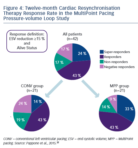
Recently, Pappone published 12-month follow-up data from this study.28,38 The trend observed at 3 months was sustained; LVESV and EF were significantly improved in the MultiPoint pacing group relative to the CONV group. A clinical benefit was also seen: improvement of more than two NYHA classes was seen in a much higher proportion of MultiPoint pacing patients compared with conventional pacing. According to the definition of response (ESV reduction of ≥15 % and alive status at 12 months), 67 % of the overall patient population were responders, with a 76 % responder rate in the MultiPoint pacing group compared with 57 % in the CONV group. This was further subdivided into superresponders (ESV reduction ≥30 %), responders (ESV reduction ≥15 and <30 %), non-responders (ESV reduction ≥0 and <15 %) and negative responders (ESV increase). The MultiPoint pacing group showed a lower rate of negative responders (10 versus 25 %) and a higher rate of super responders (33 versus 14 %) compared with the CONV group (see Figure 4).38 An example of acute haemodynamic measurements and echo measurements were given in a super responder: these included a decrease in EDV from 310 ml at baseline to 235 ml at 3 months and 211 ml at 12 months. The decrease in EF was 35 % at baseline and 41 % at both 3 and 12 months.38

Dr Calovic next posed the question: can MultiPoint pacing improve the response in patients receiving conventional CRT? At CRT implant programmed with simultaneous biventricular pacing, patients were assessed by an echo exam (LVESV, LVEDV, LVEF) as well as undergoing NYHA class assessment. At 12 months, patients underwent follow-up assessments and CRT programming was switched to MultiPoint pacing. Further follow-up assessments were performed at 16 months. Following the switch, the two non-responders to conventional CRT both became responders with MultiPoint pacing while experiencing an additional reduction in ESV of -33 % and -20 % and an improvement in EF of +15 % and +4 % at 16 months relative to the 12-month exam. Five of the six responders to conventional CRT remained responders with MultiPoint pacing, with four patients experiencing additional ESV change of at least -10 %. In terms of NYHA class changes, all patients were Class II pre-implant, all improved to Class II at 12 months and after 4 months of MultiPoint pacing, four patients improved to Class I while the remaining five patients remained in Class II.39

Twelve-month Cardiac Resynchronisation Therapy Response Rate in the MultiPoint Pacing Pressure-volume Loop Study

Article image

In conclusion, MultiPoint LV pacing in a single coronary sinus branch results in significant improvements in acute haemodynamic function and long-term CRT response relative to conventional biventricular pacing. Non-responders to conventional CRT may benefit from activating MultiPoint pacing and may be converted to responders. In addition, CRT responders may experience additional LV remodeling and/or increased LV function beyond that received from the conventional therapy.

References

  1. Epstein AE, DiMarco JP, Ellenbogen KA, et al. 2012 ACCF/AHA/ HRS focused update incorporated into the ACCF/AHA/HRS 2008 guidelines for device-based therapy of cardiac rhythm abnormalities: a report of the American College of Cardiology Foundation/American Heart Association Task Force on Practice Guidelines and the Heart Rhythm Society. J Am Coll Cardiol 2013;61:e6–75.
  2. Bristow MR, Saxon LA, Boehmer J, et al. Cardiacresynchronization therapy with or without an implantable defibrillator in advanced chronic heart failure. N Engl J Med 2004;350:2140–50.
  3. Cleland JG, Daubert JC, Erdmann E, et al. The effect of cardiac resynchronization on morbidity and mortality in heart failure. N Engl J Med 2005;352:1539–49.
  4. Moss AJ, Hall WJ, Cannom DS, et al. Cardiac-resynchronization therapy for the prevention of heart-failure events. N Engl J Med 2009;361:1329–38.
  5. Curtis AB, Yancy CW, Albert NM, et al. Cardiac resynchronization therapy utilization for heart failure: findings from IMPROVE HF. Am Heart J 2009;158:956–64.
  6. Perrin MJ, Green MS, Redpath CJ, et al. Greater response to cardiac resynchronization therapy in patients with true complete left bundle branch block: a PREDICT substudy. Europace 2012;14:690–5.
  7. Zareba W, Klein H, Cygankiewicz I, et al. Effectiveness of Cardiac Resynchronization Therapy by QRS Morphology in the Multicenter Automatic Defibrillator Implantation Trial- Cardiac Resynchronization Therapy (MADIT-CRT). Circulation 2011;123:1061–72.
  8. O‘Donnell D, Lin T, Swale M, et al. Long-term clinical response to cardiac resynchronisation therapy under a multidisciplinary model. Intern Med J 2013;43:1216–23.
  9. Ploux S, Eschalier R, Whinnett ZI, et al. Electrical dyssynchrony induced by biventricular pacing: implications for patient selection and therapy improvement. Heart Rhythm 2015;12:782–91.
  10. Saxon LA, Olshansky B, Volosin K, et al. Influence of left ventricular lead location on outcomes in the COMPANION study. J Cardiovasc Electrophysiol 2009;20:764–8.
  11. Singh JP, Klein HU, Huang DT, et al. Left ventricular lead position and clinical outcome in the multicenter automatic defibrillator implantation trial-cardiac resynchronization therapy (MADIT-CRT) trial. Circulation 2011;123:1159–66.
  12. Regoli F, Auricchio A. The role of invasive mapping in the electrophysiology laboratory. Europace 2009;11(Suppl. 5):v40–5.
  13. Singh JP, Fan D, Heist EK, et al. Left ventricular lead electrical delay predicts response to cardiac resynchronization therapy. Heart Rhythm 2006;3:1285–92.
  14. Zanon F, Baracca E, Pastore G, et al. Determination of the longest intrapatient left ventricular electrical delay may predict acute hemodynamic improvement in patients after cardiac resynchronization therapy. Circ Arrhythm Electrophysiol 2014;7:377–83.
  15. Gold MR, Leman RB, Wold N, et al. The effect of left ventricular electrical delay on the acute hemodynamic response with cardiac resynchronization therapy. J Cardiovasc Electrophysiol 2014;25:624–30.
  16. van Geldorp IE, Delhaas T, Hermans B, et al. Comparison of a non-invasive arterial pulse contour technique and echo Doppler aorta velocity-time integral on stroke volume changes in optimization of cardiac resynchronization therapy. Europace 2011;13:87–95.
  17. Lane RE, Chow AW, Mayet J, et al. The interaction of interventricular pacing intervals and left ventricular lead position during temporary biventricular pacing evaluated by tissue Doppler imaging. Heart 2007;93:1426–32.
  18. Gold MR, Birgersdotter-Green U, Singh JP, et al. The relationship between ventricular electrical delay and left ventricular remodelling with cardiac resynchronization therapy. Eur Heart J 2011;32:2516–24.
  19. Kandala J, Upadhyay GA, Altman RK, et al. QRS morphology, left ventricular lead location, and clinical outcome in patients receiving cardiac resynchronization therapy. Eur Heart J 2013;34:2252–62.
  20. Khan FZ, Virdee MS, Palmer CR, et al. Targeted left ventricular lead placement to guide cardiac resynchronization therapy: the TARGET study: a randomized, controlled trial. J Am Coll Cardiol 2012;59:1509–18.
  21. Lin T, Crosby P, Sugumar H, et al. Implant electrical characteristics predict response to cardiac resynchronization therapy, WJCD, 2014;4:513–21.
  22. Mafi Rad M, Blaauw Y, Debie L, et al. Evaluation of left ventricular endocardial cardiac resynchronization therapy in a non-responder with ventricular arrhythmias, Indian Pacing Electrophysiol J, 2014;14:32–6.
  23. Leclercq C, Gadler F, Kranig W, et al. A randomized comparison of triple-site versus dual-site ventricular stimulation in patients with congestive heart failure. J Am Coll Cardiol 2008;51:1455–62.
  24. Thibault B, Dubuc M, Khairy P, et al. Acute haemodynamic comparison of multisite and biventricular pacing with a quadripolar left ventricular lead. Europace 2013;15:984–91.
  25. Rinaldi CA, Leclercq C, Kranig W, et al. Improvement in acute contractility and hemodynamics with multipoint pacing via a left ventricular quadripolar pacing lead. J Interv Card Electrophysiol 2014;40:75–80.
  26. Pappone C, Calovic Z, Vicedomini G, et al. Multipoint left ventricular pacing improves acute hemodynamic response assessed with pressure-volume loops in cardiac resynchronization therapy patients. Heart Rhythm 2014;11:394–401.
  27. Pappone C, Calovic Z, Vicedomini G, et al. Multipoint left ventricular pacing in a single coronary sinus branch improves mid-term echocardiographic and clinical response to cardiac resynchronization therapy. J Cardiovasc Electrophysiol 2015;26:58–63.
  28. Zanon F, Baracca E, Pastore G, et al. Multipoint pacing by a left ventricular quadripolar lead improves the acute hemodynamic response to CRT compared with conventional biventricular pacing at any site. Heart Rhythm 2015;12:975–81.
  29. Forleo GB, Santini L, Potenza D, et al. Impact of multi-point left ventricular pacing on QRS duration and left ventricular ejection fraction. Preliminary results from a multicenter prospective study. Presented at HRS, Boston, May 2015 Abstract no p004-183.
  30. Fornwalt BK, Sprague WW, BeDell P, et al. Agreement is poor among current criteria used to define response to cardiac resynchronization therapy. Circulation 2010;121:1985–91.
  31. Abraham WT. Results from the FREEDOM Trial – Assess the Safety and Efficacy of Frequent Optimization of Cardiac Resynchronization Therapy. SP08. Late-Breaking Clinical Trials, HRS 2010. Denver, Colorado, US.
  32. Ellenbogen KA, Gold MR, Meyer TE, et al. Primary results from the SmartDelay determined AV optimization: a comparison to other AV delay methods used in cardiac resynchronization therapy (SMART-AV) trial: a randomized trial comparing empirical, echocardiography-guided, and algorithmic atrioventricular delay programming in cardiac resynchronization therapy. Circulation 2010;122:2660–8.
  33. Pappone C, Rosanio S, Oreto G, et al. Cardiac pacing in heart failure patients with left bundle branch block: impact of pacing site for optimizing left ventricular resynchronization. Ital Heart J 2000;1:464–9.
  34. Shetty AK, Sohal M, Chen Z, et al. A comparison of left ventricular endocardial, multisite, and multipolar epicardial cardiac resynchronization: an acute haemodynamic and electroanatomical study. Europace 2014;16:873–9.
  35. Thibault B, Dubuc M, Karst E, et al. Design of an acute dP/ dt hemodynamic measurement protocol to isolate cardiac effect of pacing. J Card Fail 2014;20:365-72.
  36. Rinaldi CA, Kranig W, Leclercq C, et al. Acute effects of multisite left ventricular pacing on mechanical dyssynchrony in patients receiving cardiac resynchronization therapy. J Card Fail 2013;19:731-8.
  37. Pappone C, Calovic Z, Vicedomini G, et al. Improving cardiac resynchronization therapy response with multipoint left ventricular pacing: Twelve-month follow-up study. Heart Rhythm 2015;12:1250–8.
  38. Pappone A, Calovic Z, Cuko A, et al. Multipoint left ventricular pacing provides additional echocardiographic benefit to responders and non-responders to conventional cardiac resynchronization therapy. Eur Heart J 2015; Suppl A: A12-A17.