Article

Mark E Josephson: Clinical Investigator

Register or Login to View PDF Permissions
Permissions× For commercial reprint enquiries please contact Springer Healthcare: ReprintsWarehouse@springernature.com.

For permissions and non-commercial reprint enquiries, please visit Copyright.com to start a request.

For author reprints, please email rob.barclay@radcliffe-group.com.
Average (ratings)
No ratings
Your rating

Abstract

Mark E Josephson entered the world of clinical cardiac electrophysiology (EP) almost at its inception (1972); with so much to learn and so many directions one could take, he dived into the field with unbridled enthusiasm and an uncommon – perhaps almost unique – aptitude for asking questions and finding ways to answer them. Few aspects of EP escaped his indelible influence. In this short paper, I will attempt to touch on some of the high points of his astounding career as a clinical investigator.

Copyright Statement:

The copyright in this work belongs to Radcliffe Medical Media. Only articles clearly marked with the CC BY-NC logo are published with the Creative Commons by Attribution Licence. The CC BY-NC option was not available for Radcliffe journals before 1 January 2019. Articles marked ‘Open Access’ but not marked ‘CC BY-NC’ are made freely accessible at the time of publication but are subject to standard copyright law regarding reproduction and distribution. Permission is required for reuse of this content.

The Electrocardiogram

In the entire realm of his work, Dr Josephson’s greatest love was the simple electrocardiogram (ECG). His discernment, based on the ECG, into the patient’s history and disease processes was legendary. Among the more important and insightful contributions he made to our understanding of heart disease and arrhythmias from the ECG are: observations of alternating preexcitation patterns in patients with Wolff-Parkinson-White (WPW) pattern who had more than one accessory atrioventricular (AV) accessory pathway (AP),1 and a patient with 1:2 conduction AV using an AV pathway;2 the nature of electrocardiographic ‘left atrial enlargement’;3 multiple ECG manifestations of ventricular tachycardia (VT) in the same patient;4,5 use of the 12-lead ECG to localise exit sites of post-infarction VT6,7 and premature ventricular complexes;8 studies on electrical alternans in wide complex tachycardias;9 a highly useful method of distinguishing VT from supraventricular tachycardia (SVT) with aberration in left bundle branch block (LBBB) wide-QRS tachycardias;10 variations in the ECG expression of atrial flutter;11 differentiation of insertion sites of atriofascicular pathways;12 classification of which portion of which coronary artery was affected in myocardial infarction;13,14 features of the ECG in what appears to be right ventricular outflow tract ectopy and VT that correlate with difficult catheter ablation;15 distinction of the causes of T-wave inversion (post-pacing cardiac memory versus ischaemia),16 and whether LBBB was new or old (with implications for patients with chest pain and LBBB on ECG).17

Throughout his career, Dr Josephson gave courses on ECG interpretation with his close friend Prof Hein Wellens; most EP fellows of the last 25 years have attended these legendary courses and come away with a new appreciation of this old tool and how much information can be extracted by masters such as Josephson and Wellens. In the later years of his career, he was invited along with Prof Wellens to provide a regular contribution to the prestigious journal Heart Rhythm – ‘Josephson and Wellens’ ECG Lessons: A Monthly Visit to the 12-Lead ECG’. Here, they retrieved some of the choice jewels from their treasures of ECGs obtained over the prior half-century, sharing their insights into how the ECG could yield important and often surprising information about the patient and their proper diagnosis and management. These pearls bear careful reading by the serious student of electrophysiology. Josephson was often heard to state something like the following: “The ECG never lies, but it doesn’t always tell you all it knows.”

A couple of anecdotes are worth sharing here. In preparing our 1988 paper on correlation of the 12-lead ECG with ‘site of origin’ of VT, some of the ECGs had been recorded as a long continuous strip from which I had to cut several complexes of individual leads and affix them to paper in a standard 12-lead ECG format. In one case, I had inadvertently mounted a lead V5 upside down; as I was reviewing a stack of these ECGs with Dr Josephson, he immediately noted the error. I was of course very embarrassed and apologetic but he was not as bothered by it as I was, knowing what it should look like. He could also name from which patient most of the ECGs came. In another instance, during a regular Monday ECG conference in a darkened room, a fellow was squirming uncomfortably at the front of the room trying to analyse an ECG projected on the screen that showed an atrial bradycardia, incomplete right bundle branch block and mild right ventricular hypertrophy. At that moment, Dr Josephson burst into the room looking for one of the other fellows; he glanced at the ECG on the screen and blurted out the diagnosis (sinus venosus atrial septal defect) and the name of the patient (one of his). Such was his command of the ECG. Although most cardiologists interpret the ECG well, and some even interpret it expertly, Dr Josephson interpreted and understood the ECG deeply, as do but a few others.

Figure 1: Diagram Showing How Recordings are Made Pre- and Post-left Ventricular Endocardial Resection for Treatment of Ventricular Tachycardia

Article image

Supraventricular Arrhythmias

Dr Josephson started his investigative career with supraventricular arrhythmias such as SVT and atrial fibrillation (AF) and flutter. In a general overview on paroxysmal SVT18 – on which he was the sole author – he showed even this early in his career (1978) how extensive his understanding of this family of arrhythmias was (as well as how much was still unknown). Among his favourite arrhythmias was AV nodal reentrant tachycardia (AVNRT), on which he published extensively throughout his career. The first of these studies investigated whether the atrium was a necessary component in AV nodal reentry.19 Following this were investigations into upper and lower common pathways between the tachycardia circuit and atrium (above) and His bundle (below);20,21 using the ΔHA interval (HA during SVT versus HA during ventricular pacing at the SVT rate) to distinguish AV nodal reentry from orthodromic SVT using a septal AP;22 variations in retrograde conduction pathways in AVNRT, in which he showed that retrograde conduction did not necessarily have the same atrial activation pattern during SVT and retrograde conduction with ventricular pacing;23,24 the origin of accelerated junctional rhythm during slow pathway ablation for AVNRT;25 that standard slow pathway ablation of AVNRT suffices in patients with earliest retrograde activation in the coronary sinus;26 the unusual rhythm disturbance of 1:2 AV conduction during sinus rhythm coexisting with typical AV nodal reentry (which many thought impossible);27 and his final major work on AVNRT, in which he matter-of-factly corrected some of his own prior work in the light of newer evidence.28 Dr Josephson also added to knowledge and practice in a paper on selection of sites for ablation of APs in WPW, in particular using the unipolar electrogram;29 the previously-noted atriofascicular AP insertion study;12 and several papers investigating AF. These included evaluating the success of AF ablation by pulmonary vein (PV) isolation when entrance block into, and exit block out of, the PV were obtained,30,31 as well as correlating acute PV reconnection after ablation with poorer outcomes;32 a case in which AF was contained within a PV after isolation;33 and pointing out the frequency of aortic wall injury during AF ablation procedures.34 From the start of his career to its end, despite his plethora of other investigative interests, his attention was never very far from SVTs.

Figure 2: Recordings from 20-bipolar Electrode Array

Article image

Ventricular Tachycardia

The area of clinical investigation for which Dr Josephson is most renowned is VT. In the 1970s, post-infarction VT was a major problem without a good solution. Antiarrhythmic drugs were palliative at best, and neither catheter ablation nor implantable defibrillators had been developed. Dr Josephson believed that effective treatment might hinge on a better understanding of the nature of the arrhythmia – which at that time was poor. He and his colleagues began a series of investigations that not only changed many aspects of the care of patients with VT, but eventually those with many other types of arrhythmias (to which the principles learned in VT investigation could be applied). The first studies he published in this area were an astounding series of four landmark papers, appearing in Circulation over a span of 13 months.4,35–37 This was a planned series (the first is entitled ‘Recurrent Sustained Ventricular Tachycardia. 1. Mechanisms’) setting forth the state of the art at that time, and showing that:

  • VT in this setting was due to reentry;
  • Reentry occurred on the endocardium;
  • Reentry did not involve the proximal His-Purkinje system; and
  • Reentry occurred in a relatively small area.

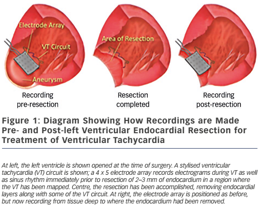
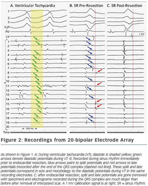
These findings led directly to surgical treatment of VT by excising endocardial tissue proven to be essential for ongoing arrhythmia; finally, a cure for VT was possible.38,39 A series of papers followed this providing further insights into the behaviour of VT5,40,41 – where circuits were, patterns of activation during VT, what accounted for multiple QRS morphologies of VT, observations about ‘dead-end’ conduction pathways with 2:1 conduction during VT – and led to innovations in technique, resulting in improved outcomes.42 This, in turn, paved the way for subsequent work in catheter ablation of VT, in which Dr Josephson was an active investigator.43 Additional investigations involved the aforementioned use of the ECG during VT to localise exit sites from circuits,6,7 pace-mapping to corroborate activation mapping;44,45 and one of his favourite studies, the effect of endocardial tissue resection on electrograms at sites of reentry.46 In this study, a multipolar electrode array was positioned on a portion of endocardium to which VT had been mapped during the procedure; recordings were made during sinus rhythm (and usually also VT); the endocardium was then removed by undermining with scissors; and finally, the array was placed back in the same position where recordings were repeated during sinus rhythm (see Figure 1). This showed that endocardial late potentials (recorded after the end of the QRS complex) that correlated with mid-diastolic potentials during VT were located on the endocardial surface (present prior to resection, absent after), and that the larger signals seen during the QRS complex were recorded from deeper layers, with their amplitudes decreased by endocardial scar (see Figure 2). He also participated in studies of VT in the absence of structural heart disease47 as well as in the setting of congenital heart disease (repaired tetralogy of Fallot, Ebstein anomaly).48,49 One of his last battles against VT was in the form of prophylactic catheter ablation of VT to try to prevent shocks from implanted defibrillators;50 he always believed prevention was a better strategy than rescue (analogy: preventing injury by preventing a motor vehicle accident, versus rescue by airbag), so this study was a natural fit for him.

Mechanisms and Tools of Investigation

Dr Josephson was known for his earnest search for a more thorough understanding of the mechanisms of arrhythmias, strongly believing that correct therapy depended on knowing the correct diagnosis. While this seems self-evident, some electrophysiologists still perform catheter ablation procedures based on a presumption of a diagnosis and mechanism (reentry versus automaticity, for instance), rather than taking the extra few seconds or minutes needed to prove the case. A long and ultimately unsuccessful procedure can result from a strategy of presumption, if incorrect. Dr Josephson penned two perspective papers entitled ‘Electrophysiology at a Crossroads’51,52 that set forth his reservations about how EP practices had evolved from mechanismand evidence-based care to what was expedient and economically driven; these are required reading in many training programmes.

Dr Josephson’s research into mechanisms of arrhythmias was spread throughout his career, starting with a publication on the effects of lidocaine in man.53 A few years later, the previously-mentioned work on mechanisms of VT appeared as the first of many regarding this arrhythmia. Following this were papers describing continuous electrical activity during VT;54 the mechanism of ventricular fibrillation;55 structure and ultrastructure of surgically-removed specimens from VT patients;56 and the meaning of endocardial late potentials.57 A series of monumental papers on resetting and entrainment of arrhythmias began in 198558 and continued for a decade thereafter, including resetting response patterns;59,60 resetting in the presence of fusion;61 comparing results of single-beat resetting versus entrainment;62 predictability of overdrive pacing to terminate VT;63 and investigation as to whether functional or fixed lines of conduction block are present in VT.64 The lessons learned from these studies provide substantial help in some cases of complex reentry, which are increasingly encountered after ablation of AF. Dr Josephson provided a lucid explanation of the mechanism of the rare but important disorder known as paroxysmal AV block.65 He also contributed to the development of the first electroanatomical mapping system.66

Dr Josephson participated in investigations into drug therapy of arrhythmias and the adverse effects of these drugs; cardiovascular genetics; animal models of arrhythmias; heart failure; valvular heart disease; device-based therapy of atrial and ventricular arrhythmias; clinical trials in arrhythmia therapy; and healthcare economics.

Conclusion

The panoply of contributions by Dr Josephson to our understanding of cardiac arrhythmias is far too broad to recount in a short paper. Even in failing health in his last two years here, he continued to develop new ideas about ways to investigate arrhythmias. His contributions to the field are large, both in number and impact; practically every procedure that a practicing electrophysiologist does on any given day has been influenced by Dr Josephson’s work. These accomplishments were of course important to him; far more important were his relationships with his family, colleagues and trainees. Ever the advocate and friend, he never saw himself as too important or busy to help someone understand a difficult point, or offer patient care or career advice when asked. A good clinical investigator does good work; an exceptional investigator inspires those with whom he works to launch out onto their own investigative paths. Dr Josephson was clearly among the exceptional. Thus it is that, through his students – who he taught how to think and ask questions – his work continues.

Lastly, although Dr Josephson made many new discoveries and devised new therapies in his investigative career, some of the conclusions he made from these studies turned out to be less than completely correct; he was big enough to own up to these and criticise his own prior work. A pioneer in any field, like an explorer in new territory, occasionally goes down a seemingly promising path that eventually leads nowhere. Far more often than not, however, Dr Josephson led us down a path of new discovery and we find, in looking back, that the ‘hits’ far outnumbered the ‘misses’. It is difficult to imagine where our knowledge about arrhythmias would be, and how we would be practicing EP today, were it not for the contributions of this one man. His legacy as a pioneer in our field is secure.

References

  1. Josephson ME, Caracta AR, Lau SH. Alternating type A and type B Wolff-Parkinson-White syndrome. Am Heart J 1974;87:363–6.
    Crossref | PubMed
  2. Josephson ME, Seides SF, Damato AN. Wolff-Parkinson- White syndrome with 1:2 atrioventricular conduction. Am J Cardiol 1976;37:1094–6.
    Crossref | PubMed
  3. Josephson ME, Kastor JA, Morganroth J. Electrocardiographic left atrial enlargement. Electrophysiologic, echocardiographic and hemodynamic correlates. Am J Cardiol 1977;39:967–71.
    Crossref | PubMed
  4. Josephson ME, Horowitz LN, Farshidi A, et al. Recurrent sustained ventricular tachycardia. 4. Pleomorphism. Circulation 1979;59:459–68.
    Crossref | PubMed
  5. Miller JM, Kienzle MG, Harken AH, Josephson ME. Morphologically distinct sustained ventricular tachycardias in coronary artery disease: significance and surgical results. J Am Coll Cardiol 1984;4:107–39. 
    Crossref | PubMed
  6. Josephson ME, Horowitz LN, Waxman HL, et al. Sustained ventricular tachycardia: role of the 12-lead electrocardiogram in localizing site of origin. Circulation 1981;64:257–72.
    Crossref | PubMed
  7. Miller JM, Marchlinski FE, Buxton AE, Josephson ME. Relationship between the 12-lead electrocardiogram during ventricular tachycardia and endocardial site of origin in patients with coronary artery disease. Circulation 1988;77:759– 66.
    Crossref | PubMed
  8. Josephson ME. The origin of premature ventricular complexes– role and limitations of the 12-lead electrocardiogram. Int J Cardiol 1982;2:87–90.
    Crossref | PubMed
  9. Kremers MS, Miller JM, Josephson ME. Electrical alternans in wide complex tachycardias. Am J Cardiol 1985;56:305–8.
    Crossref | PubMed
  10. Kindwall KE, Brown J, Josephson ME. Electrocardiographic criteria for ventricular tachycardia in wide complex left bundle branch block morphology tachycardias. Am J Cardiol 1988;61:1279–83.
    Crossref | PubMed
  11. Milliez P, Richardson AW, Obioha-Ngwu O, et al. Variable electrocardiographic characteristics of isthmus-dependent atrial flutter. J Am Coll Cardiol 2002;40:1125–32.
    Crossref | PubMed
  12. Gandhavadi M, Sternick EB, Jackman WM, et al. Characterization of the distal insertion of atriofascicular accessory pathways and mechanisms of QRS patterns in atriofascicular antidromic tachycardia. Heart Rhythm 2013;10:1385–92.
    Crossref | PubMed
  13. Zimetbaum PJ, Krishnan S, Gold A, et al. Usefulness of ST-segment elevation in lead III exceeding that of lead II for identifying the location of the totally occluded coronary artery in inferior wall myocardial infarction. Am J Cardiol 1998;81:918–9.
    Crossref | PubMed
  14. Zimetbaum PJ, Josephson ME. Use of the electrocardiogram in acute myocardial infarction. N Engl J Med 2003;348:933–40.
    Crossref | PubMed
  15. Hachiya H, Hirao K, Sasaki T, et al. Novel ECG predictor of difficult cases of outflow tract ventricular tachycardia: peak deflection index on an inferior lead. Circ J 2010;74:256–61.
    Crossref | PubMed
  16. Shvilkin A, Ho KK, Rosen MR, Josephson ME. T-vector direction differentiates postpacing from ischemic T-wave inversion in precordial leads. Circulation 2005;111:969–74.
    Crossref | PubMed
  17. Shvilkin A, Bojovic B, Vajdic B, et al. Vectorcardiographic and electrocardiographic criteria to distinguish new and old left bundle branch block. Heart Rhythm 2010;7:1085–92.
    Crossref | PubMed
  18. Josephson ME. Paroxysmal supraventricular tachycardia: an electrophysiologic approach. Am J Cardiol 1978;41:1123–6.
    Crossref | PubMed
  19. Josephson ME, Kastor JA. Paroxysmal supraventricular tachycardia: is the atrium a necessary link? Circulation 1976;54:430–5.
    Crossref | PubMed
  20. Miller JM, Rosenthal ME, Vassallo JA, Josephson ME. Atrioventricular nodal reentrant tachycardia: studies on upper and lower 'common pathways'. Circulation 1987;75:930–40.
    Crossref | PubMed
  21. Anselme F, Poty H, Cribier A, et al. Entrainment of typical AV nodal reentrant tachycardia using para-Hisian pacing: evidence for a lower common pathway within the AV node. J Cardiovasc Electrophysiol 1999;10:655–61.
    Crossref | PubMed
  22. Miller JM, Rosenthal ME, Gottlieb CD, et al. Usefulness of the delta HA interval to accurately distinguish atrioventricular nodal reentry from orthodromic septal bypass tract tachycardias. Am J Cardiol 1991;68:1037–44.
    Crossref | PubMed
  23. Anselme F, Hook B, Monahan K, et al. Heterogeneity of retrograde fast-pathway conduction pattern in patients with atrioventricular nodal reentry tachycardia: observations by simultaneous multisite catheter mapping of Koch's triangle. Circulation 1996;93:960–8.
    Crossref | PubMed
  24. Anselme F, Papageorgiou P, Monahan K, et al. Presence and significance of the left atrionodal connection during atrioventricular nodal reentrant tachycardia. Am J Cardiol 1999;83:1530–6.
    Crossref | PubMed
  25. Boyle NG, Anselme F, Monahan K, et al. Origin of junctional rhythm during radiofrequency ablation of atrioventricular nodal reentrant tachycardia in patients without structural heart disease. Am J Cardiol 1997;80:575–80.
    Crossref | PubMed
  26. Chen J, Anselme F, Smith TW, et al. Standard right atrial ablation is effective for atrioventricular nodal reentry with earliest activation in the coronary sinus. J Cardiovasc Electrophysiol 2004;15:2–7.
    Crossref | PubMed
  27. Germano JJ, Essebag V, Papageorgiou P, Josephson ME. Concealed and manifest 1:2 tachycardia and atrioventricular nodal reentrant tachycardia: manifestations of dual atrioventricular nodal physiology. Heart Rhythm 2005;2:536–9.
    Crossref | PubMed
  28. Katritsis DG, Josephson ME. Classification of electrophysiological types of atrioventricular nodal re-entrant tachycardia: a reappraisal. Europace 2013;15:1231–40.
    Crossref | PubMed
  29. Grimm W, Miller J, Josephson ME. Successful and unsuccessful sites of radiofrequency catheter ablation of accessory atrioventricular connections. Am Heart J 1994;128:77–87.
    Crossref | PubMed
  30. Essebag V, Baldessin F, Reynolds MR, et al. Non-inducibility post-pulmonary vein isolation achieving exit block predicts freedom from atrial fibrillation. Eur Heart J 2005;26:2550–5. PMCID:PMC1361286
    Crossref | PubMed
  31. Essebag V, Wylie JV Jr, Reynolds MR, et al. Bi-directional electrical pulmonary vein isolation as an endpoint for ablation of paroxysmal atrial fibrillation. J Interv Card Electrophysiol 2006;17:111–7.; PMCID:PMC1937574
    Crossref | PubMed
  32. Anter E, Contreras-Valdes FM, Shvilkin A, et al. Acute pulmonary vein reconnection is a predictor of atrial fibrillation recurrence following pulmonary vein isolation. J Interv Card Electrophysiol 2014;39:225–32.
    Crossref | PubMed
  33. Josephson ME. Isolation of atrial fibrillation to a pulmonary vein. Europace 2003;5:365–6.
    Crossref | PubMed
  34. Tung P, Hong SN, Chan RH, et al. Aortic injury is common following pulmonary vein isolation. Heart Rhythm 2013;10:653–8.
    Crossref | PubMed
  35. Horowitz LN, Josephson ME, Farshidi A, et al. Recurrent sustained ventricular tachycardia 3. Role of the electrophysiologic study in selection of antiarrhythmic regimens. Circulation 1978;58:986–97.
    Crossref | PubMed
  36. Josephson ME, Horowitz LN, Farshidi A, Kastor JA. Recurrent sustained ventricular tachycardia. 1. Mechanisms. Circulation 1978;57:431–40.
    Crossref | PubMed
  37. Josephson ME, Horowitz LN, Farshidi A, et al. Recurrent sustained ventricular tachycardia. 2. Endocardial mapping. Circulation 1978;57:440–7.
    Crossref | PubMed
  38. Harken AH, Josephson ME, Horowitz LN. Surgical endocardial resection for the treatment of malignant ventricular tachycardia. Ann Surg 1979;190:456–60. PMCID:PMC1344508
    Crossref | PubMed
  39. Josephson ME, Harken AH, Horowitz LN. Endocardial excision: a new surgical technique for the treatment of recurrent ventricular tachycardia. Circulation 1979;60:1430–9.
    Crossref | PubMed
  40. Miller JM, Kienzle MG, Harken AH, Josephson ME. Subendocardial resection for ventricular tachycardia: predictors of surgical success. Circulation 1984;70:624–31.
    Crossref | PubMed
  41. Miller JM, Vassallo JA, Hargrove WC, Josephson ME. Intermittent failure of local conduction during ventricular tachycardia. Circulation 1985;72:1286–92. 
    Crossref | PubMed
  42. Hargrove WC, 3rd, Miller JM, Vassallo JA, Josephson ME. Improved results in the operative management of ventricular tachycardia related to inferior wall infarction. Importance of the annular isthmus. J Thorac Cardiovasc Surg 1986;92:726–32.
    Crossref | PubMed
  43. El-Shalakany A, Hadjis T, Papageorgiou P, et al. Entrainment/ mapping criteria for the prediction of termination of ventricular tachycardia by single radiofrequency lesion in patients with coronary artery disease. Circulation 1999;99:2283–9.
    Crossref | PubMed
  44. Josephson ME, Waxman HL, Cain ME, et al. Ventricular activation during ventricular endocardial pacing. II. Role of pace-mapping to localize origin of ventricular tachycardia. Am J Cardiol 1982;50:11–22.
    Crossref | PubMed
  45. Waxman HL, Josephson ME. Ventricular activation during ventricular endocardial pacing: I. Electrocardiographic patterns related to the site of pacing. Am J Cardiol 1982;50:1–10.
    Crossref | PubMed
  46. Miller JM, Tyson GS, Hargrove WC 3rd, et al. Effect of subendocardial resection on sinus rhythm endocardial electrogram abnormalities. Circulation 1995;91:2385–91.
    Crossref | PubMed
  47. Buxton AE, Marchlinski FE, Doherty JU, et al. Repetitive, monomorphic ventricular tachycardia: clinical and electrophysiologic characteristics in patients with and patients without organic heart disease. Am J Cardiol 1984;54:997–1002.
    Crossref | PubMed
  48. Harken AH, Horowitz LN, Josephson ME. Surgical correction of recurrent sustained ventricular tachycardia following complete repair of tetralogy of Fallot. J Thorac Cardiovasc Surg 1980;80:779–81.
    PubMed
  49. Obioha-Ngwu O, Milliez P, Richardson A, et al. Ventricular tachycardia in Ebstein's Anomaly. Circulation 2001;104:E92–4.
    Crossref | PubMed
  50. Reddy VY, Reynolds MR, Neuzil P, et al. Prophylactic catheter ablation for the prevention of defibrillator therapy. N Engl J Med 2007;357:2657–65.PMCID:PMC2390777
    Crossref | PubMed
  51. Josephson ME. Electrophysiology at a crossroads. Heart Rhythm 2007;4:658–61.
    Crossref | PubMed
  52. Josephson ME. Electrophysiology at a crossroads: A revisit. Heart Rhythm 2016;13:2317–22.
    Crossref | PubMed
  53. Josephson ME, Caracta AR, Lau SH, et al. Effects of lidocaine on refractory periods in man. Am Heart J 1972;84:778–86.
    Crossref | PubMed
  54. Josephson ME, Horowitz LN, Farshidi A. Continuous local electrical activity. A mechanism of recurrent ventricular tachycardia. Circulation 1978;57:659–65.
    Crossref | PubMed
  55. Josephson ME, Spielman SR, Greenspan AM, Horowitz LN. Mechanism of ventricular fibrillation in man. Observations based on electrode catheter recordings. Am J Cardiol 1979;44:623–31.
    Crossref | PubMed
  56. Fenoglio JJ Jr, Pham TD, Harken AH, et al. Recurrent sustained ventricular tachycardia: structure and ultrastructure of subendocardial regions in which tachycardia originates. Circulation 1983;68:518–33.
    Crossref | PubMed
  57. Vassallo JA, Cassidy D, Simson MB, et al. Relation of late potentials to site of origin of ventricular tachycardia associated with coronary heart disease. Am J Cardiol 1985;55:985–9.
    Crossref | PubMed
  58. Almendral JM, Gottlieb C, Marchlinski FE, et al. Entrainment of ventricular tachycardia by atrial depolarizations. Am J Cardiol 1985;56:298–304.
    Crossref | PubMed
  59. Almendral JM, Rosenthal ME, Stamato NJ, et al. Analysis of the resetting phenomenon in sustained uniform ventricular tachycardia: incidence and relation to termination. J Am Coll Cardiol 1986;8:294–300.
    Crossref | PubMed
  60. Almendral JM, Stamato NJ, Rosenthal ME, et al. Resetting response patterns during sustained ventricular tachycardia: relationship to the excitable gap. Circulation 1986;74:722–30.
    Crossref | PubMed
  61. Rosenthal ME, Stamato NJ, Almendral JM, et al. Resetting of ventricular tachycardia with electrocardiographic fusion: incidence and significance. Circulation 1988;77:581–8.
    Crossref | PubMed
  62. Callans DJ, Hook BG, Josephson ME. Comparison of resetting and entrainment of uniform sustained ventricular tachycardia. Further insights into the characteristics of the excitable gap. Circulation 1993;87:1229–38.
    Crossref | PubMed
  63. Callans DJ, Hook BG, Mitra RL, Josephson ME. Characterization of return cycle responses predictive of successful pacingmediated termination of ventricular tachycardia. J Am Coll Cardiol 1995;25:47–53.
    Crossref | PubMed
  64. Callans DJ, Zardini M, Gottlieb CD, Josephson ME. The variable contribution of functional and anatomic barriers in human ventricular tachycardia: an analysis with resetting from two sites. J Am Coll Cardiol 1996;27:1106–11.
    Crossref | PubMed
  65. Lee S, Wellens HJ, Josephson ME. Paroxysmal atrioventricular block. Heart Rhythm 2009;6:1229–34.
    Crossref | PubMed
  66. Ben-Haim SA, Osadchy D, Schuster I, et al. Nonfluoroscopic, in vivo navigation and mapping technology. Nat Med 1996;2:1393–5.
    Crossref | PubMed