Article

Session 6: Interaction Between Underlying Atrial Tissue and Lesion

Register or Login to View PDF Permissions
Permissions× For commercial reprint enquiries please contact Springer Healthcare: ReprintsWarehouse@springernature.com.

For permissions and non-commercial reprint enquiries, please visit Copyright.com to start a request.

For author reprints, please email rob.barclay@radcliffe-group.com.
Information image
Average (ratings)
No ratings
Your rating
Copyright Statement:

The copyright in this work belongs to Radcliffe Medical Media. Only articles clearly marked with the CC BY-NC logo are published with the Creative Commons by Attribution Licence. The CC BY-NC option was not available for Radcliffe journals before 1 January 2019. Articles marked ‘Open Access’ but not marked ‘CC BY-NC’ are made freely accessible at the time of publication but are subject to standard copyright law regarding reproduction and distribution. Permission is required for reuse of this content.

The first session of Day 2 focused on the importance of the target atrial tissue as opposed to the ablation energy of the tool, because wall thickness and tissue composition play almost an important a role as the energy. Presentations described lesion efficacy and disease substrate, information derived from electrograms, and innovative approaches to speeding of ablation times.

Atrial Wall Thickness: The Missing Ingredient for Effective Tailored Ablation

Left atrial wall thickness varies with atrial location, pathological state and age. It is a potential early marker of adverse atrial remodelling and the atrial wall is the primary ablation target, making wall thickness a relevant consideration in RF ablation success, said Prof Mark O’Neill from London, UK, opening Day 2.

Based on the available literature, the thickness of the atrial wall may have a role to play in perpetuation of AF. In limited studies of patients with AF recurrence, the atrial wall has been demonstrated to be thicker in some zones than in patients without recurrence, and as the degree of fibrosis (as estimated by voltage) increases, so too does atrial wall thickness. In addition, many studies of RF lesion formation show that lesion depth is at least 3–4 mm, sometimes 4–6 mm at powers, contact force and durations typically employed for AF ablation.41–45 The atrial wall may be 2–3 mm thick, and this must be taken into consideration.

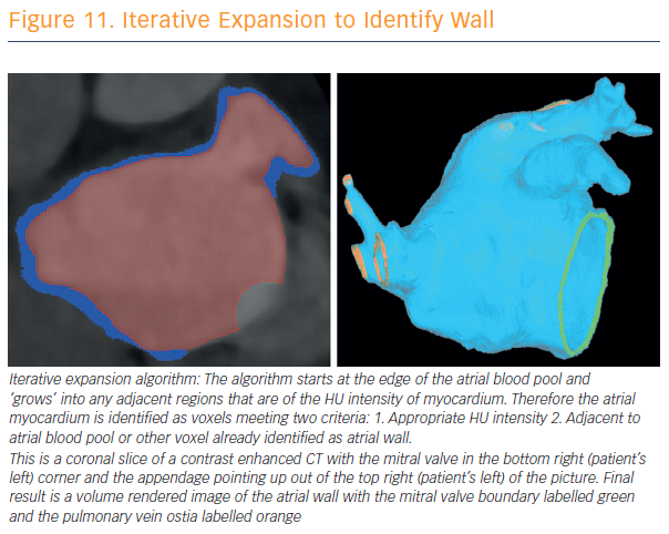
To assess and validate atrial wall thickness, 3D atrial wall thickness maps were created to inform catheter ablation procedures for AF (see Figure 11). Prof O’Neill and his team have validated their method for measurements in porcine hearts.46 CT is currently the best non-invasive modality for wall-thickness assessment, a calculation that may provide a reproducible means for assessment of disease progression. Awareness of local wall thickness is likely to encourage less rather than more ablation in most locations.

“We have been able to demonstrate that the atrial wall thickness pertaining to the appendage is greater at the anterior aspect of the appendage followed by the superior, inferior and posterior aspects,” said Prof O’Neill.

The sites of reconnection are most commonly at the thickest site, the anterior site, followed by the next thickest, and so on. Where there was acute reconnection, this was more commonly seen in thicker than thinner atrial sites. More work is needed before recommending ablation targets identified using MR or CT-derived wall thickness information. There’s also a new area to explore. It is possible that an increase in left atrial wall thickness may in fact be an early marker of propensity to AF – earlier than fibrosis detected on an MRI scan – and something that could be more widely applicable to early detection of patients at risk of AF.

Iterative Expansion to Identify Wall

Article image

Bipolar and Unipolar Electrogram Changes as In Vivo Marker for Transmural Lesion

Dr Agustín Bortone of Service de Cardiologie Hôpital Privé les Franciscaines in Nîmes, France, spoke on unipolar and bipolar electrogram changes as in vivo markers of transmurality creation. Several publications have shown that local electrogram amplitude reduction greater than 50 % is associated with the formation of transmural lesions (TL).47,48 However, the monitored bipolar (most commonly) electrogram from the tip to the first recording ring (tipring) extends beyond ablated tissues, reducing the effect of RF application on the monitored electrogram. In addition, other studies do not confirm the relationship between a halving of the electrogram amplitude and the creation of TL. Some show that achievement of split potentials rather than electrogram amplitude reduction is predictive of TL, while others argue that a signal amplitude reduction of 80% or even 90% is a marker of TL, rather than 50 %.49-51

Dr Bortone said that it seems logical to consider that the decrease in the amplitude of electrogram – either unipolar or bipolar – is correlated with the depth of the lesion and thus to TL achievement. However, with conventional catheters the electrogram analysed takes account of information that exceeds the treated tissue. There is a lack of specificity, and the percentage of exact reduction of the electrogram amplitude defining a TL is not clear.

Dr Bortone discussed and summarised the literature regarding these issues, and his analysis concluded that bipolar electrogram (BE) and unipolar electrogram amplitude reduction monitored by conventional catheters does not allow accurate TL creation assessment, in part because the perfect percentage reduction value is unknown. Bipolar electrogram amplitude reduction monitored by mini electrodes appears promising as a marker of TL creation. Unipolar electrogram morphologic changes to complete a positive signal are clear, reproducible and accurate markers of TL creation. BE morphologic changes are highly variable and, therefore, very difficult to use as real-time markers of TL creation.52 Transmurality is either functional and reversible, or corresponds to a necrotic and irreversible state.

Prof Mark O’Neill

Article image

In conclusion, the association of the monitored BE reduction (plateau) monitored by mini electrodes and the analysis of the morphologic changes of the unipolar electrogram may improve our ability to assess TL creation. The CF-sensing technology, as well as short-duration and high-power RF applications may increase the probability of creating transmural and necrotic (irreversible) lesions. The whole challenge lies in creating transmural, irreversible AND non-extramural lesions.

Can Short-duration and High-power RF Delivery be Safe and Effective?

Dr Elad Anter of Beth Israel Deaconess Medical Center in Boston, US, asked why the incidence of PV reconnection remains high despite adequate energy and tissue CF, and speculated that inadequate catheter stability, especially over a relatively long duration of 20 to 30 seconds may play a role by not necessarily creating tissue oedema. The answer may lie in higher energy for a shorter duration, which could provide more effective energy and, potentially reduced collateral injury.

Summarising numerous experiments in a thigh muscle preparation, Dr Anter said: “The combination of 90 W for 4 seconds at 10–20 g appeared to be effective and safe. These are the parameters that we have taken to the beating heart.”

He has focused on applying these findings using the QDot Micro Thermocool Smarttouch SF. Dr Anter has found that high power and short duration – 90 W and 4 seconds – of ablation can be effective in atrial tissue. It produces full-thickness, irreversible cellular damage that, in all PVs, including the left PV, are wider and more contiguous, compared to standard energy delivery. The safety of this technology requires real-time temperature monitoring, limited to 65˚C, plus an advanced irrigation technology. Collateral damage appears to be smaller compared to conventional ablation, probably due to reduced conductive heating.

Multimodality Assessment of the Atrial Fibrillation Substrate: Fat, Fractionation and LGE

Dr Saman Nazarian from the University of Pennsylvania, in Philadelphia, US, explored the impact of the substrate components. He noted that epicardial fat is associated with AF. Fat is metabolically active, releases inflammatory cytokines and adipokines into the adjacent myocardium, and exhibits rich innervation from ganglionated plexi in the proximity of the PV ostia.

He and his team studied the epicardial fat distribution and examined its association with electrogram properties.53 They noted that epicardial adipose tissue is very strongly associated with bipolar voltage, and it has a regional character, meaning that where there is epicardial adipose tissue overlying that region, the bipolar voltage amplitude is decreased. The geographic distribution of fractionated electrograms also has a strong association with overlying epicardial fat.

Fibrosis is also associated with AF. There are different modes of fibrosis: reactive interstitial fibrosis that separates muscle bundles, and reparative fibrosis (final) that replaces dead cardiomyocytes.54 The presence of fibrosis interferes with electrical continuity due to non-conductive patches, slows conduction due to longer paths, alters the balance of refractory and excitability properties, and may anchor re-entry. Furthermore, due to fibroblast electrical coupling with cardiomyocytes, fibrosis may promote ectopic activity.

“Surprisingly, right atrial fibrosis is also quite common; in fact, there’s more fibrosis in the right atrial appendage compared to the left atrial appendage. This brings into question the forgotten chamber when we’re ablating AF and the potential role of fibrosis in the right atrium,” Dr Nazarian commented.

He went on to explain his work in image-based arrhythmia substrate assessment, and its promise as a tool for arrhythmia management in the era of ‘precision medicine’.55,56 Late gadolinium enhancement (LGE) on CMR is associated with reduced voltage, decreased atrial function, and decreased conduction velocity. The association of AF persistence with AF recurrence post ablation is entirely mediated by the extent of fibrosis at baseline. However, LGE on CMR does not distinguish reactive fibrosis (i.e. potentially transient) versus replacement fibrosis, which is permanent.

“I wasn’t fully convinced of gadolinium imaging, but we started to see these beautiful linear lesions following ablation, that not only show ablation related scar but also highlight areas of pre-existing fibrosis,” said Dr Nazarian. “Atrial function also closely associates with fibrosis as measured by LGE. Passive atrial EF is closely associated with fibrosis based on our data. Active EF is just on the border of statistical significance. All strain parameters are also strongly associated with the extent of late gadolinium enhancement.” These associations have obvious implications for stroke risk stratification in the setting of AF.

References

  1. Zoni-Berisso M, Lercari F, Carazza T, Domenicucci S. Epidemiology of atrial fibrillation: European perspective. Clin Epidemiol 2014;6:213–20.
    Crossref PubMed
  2. Goette A, Kalman JM, Aguinaga L, et al. EHRA/HRS/APHRS/ SOLAECE expert consensus on atrial cardiomyopathies: Definition, characterisation, and clinical implication. J Arrhythm 2016;32:247–78.
    Crossref PubMed
  3. Wijesurendra RS, Liu A, Eichhorn C, et al. Lone atrial fibrillation is associated with impaired left ventricular energetics that persists despite successful catheter ablation. Circulation 2016;134:1068–81.
    Crossref PubMed
  4. Kolb C, Nürnbuger S, Ndrepepa G, et al. Modes of initiation of paroxysmal atrial fibrillation from analysis of spontaneously occurring episodes using a 12-lead Holter monitoring system. Am J Cardiol 2001;88:853–7.
    Crossref PubMed
  5. Ehrlich JR, Cha TJ, Zhang L, et al. Cellular electrophysiology of canine pulmonary vein cardiomyocytes: action potential and ionic current properties. J Physiol 2003;551: 801–13.
    Crossref PubMed
  6. de Groot N, van der Does L, Yaksh A, et al. Direct proof of endo-epicardial asynchrony of the atrial wall during atrial fibrillation in humans. Circ Arrhythm Electrophysiol 2016;9:pii: e003648.
    Crossref PubMed
  7. Kirchof P, Benussi S, Koetecha D, et al. 2016 ESC guidelines for the management of atrial fibrillation developed in collaboration with EACTS. Eur J Cardiothorac Surg 2016;50:e1- e88.
    Crossref PubMed
  8. Pison L, La Meir M, van Opstal A, et al. Hybrid thoracoscopic surgical and transvenous catheter ablation of atrial fibrillation. J Am Coll Cardiol 2012;60:54–61.
    Crossref PubMed
  9. Dudink E, Essers B, Holvoet W, et al. Acute cardioversion vs a wait-and-see approach for recent-onset symptomatic atrial fibrillation in the emergency department: Rationale and design of the randomized ACWAS trial. Am Heart J 2017;183:49–53.
    Crossref PubMed
  10. Ouyang F, Tilz R, Chun J, et al. Long-term results of catheter ablation in paroxysmal atrial fibrillation: Lessons from a 5-year follow-up. Circulation 2010;122:2368–77.
    Crossref PubMed
  11. Calkins H, Reynolds MR, Spector P, et al. Treatment of atrial fibrillation with antiarrhythmic drugs or radiofrequency ablation. Circ Arrythm Electrophysiol 2009;2:349–61.
    Crossref PubMed
  12. de Vos CB, Pisters R, Nieuwlaat R, et al. Progression from paroxysmal to persistent atrial fibrillation: clinical correlates and prognosis. J Am Coll Cardiol 2010;55:725–31.
    Crossref PubMed
  13. Weimar T, Schena S, Bailey MS, et al. The Cox-Maze procedure for lone atrial fibrillation: a single-center experience over 2 decades. Circ Arrhythm Electrophysiol 2012;5:8–14.
    Crossref PubMed
  14. Gallagher MM, Camm AJ. Classification of atrial fibrillation. Pacing Clin Electrophysiol 1997;20:1603–5.
    Crossref PubMed
  15. January CT, Wann LS, Alpert JS. 2014 AHA/ACC/HRS guideline for the management of patients with atrial fibrillation: executive summary: a report of the American College of Cardiology/American Heart Association Task Force on practice guidelines and the Heart Rhythm Society. Circulation 2014;130:2071–104.
    Crossref PubMed
  16. Platonov PG, Mitrofanova LB, Orshanskaya V, et al. Structural abnormalities in atrial walls are associated with presence and persistency of atrial fibrillation but not with age. J Am Coll Cardiol 2011;58:2225–32.
    Crossref PubMed
  17. Kottkamp H, Schreiber D. The substrate in “early persistent” atrial fibrillation: arrhythmia induced, risk factor induced, or from a specific fibrotic atrial cardiomyopathy? J Am Coll Cardiol Clin Electrophysiol 2016;2:140–2.
    Crossref
  18. Kottkamp H, Schreiber D, Moser F, Rieger A. Therapeutic approaches to atrial fibrillation ablation targeting atrial fibrosis. J Am Coll Cardiol EP 2017;3:643–53.
    Crossref
  19. Gianni C, Atoui M, Mohanty S, et al. Difference in thermodynamics between two types of esophageal temperature probes: Insights from an experimental study. Heart Rhythm 2016;13:2195–200.
    Crossref PubMed
  20. Rahman F, Kwan GF, Benjamin EJ. Global epidemiology of atrial fibrillation. Nat Rev Cardiol 2014;11:639–54.
    Crossref PubMed
  21. Steinberg BA, Holmes DN, Ezekowitz MD, et al. Rate versus rhythm control for management of atrial fibrillation in clinical practice: results from the Outcomes Registry for Better Informed Treatment of Atrial Fibrillation (ORBIT-AF) registry. Am Heart J 2013;165:622-9.
    Crossref PubMed
  22. Calkins H, Reynolds MR, Spector P, et al. Treatment of atrial fibrillation with antiarrhythmic drugs or radiofrequency ablation: two systematic literature reviews and metaanalyses. Circ Arrhythm Electrophysiol 2009;2:349-61.
    Crossref PubMed
  23. Medtronic internal estimates
  24. Raatikainen MJ, Arnar DO, Merkely B, et al. Access to and clinical use of cardiac implantable electronic devices and interventional electrophysiological procedures in the European Society of Cardiology Countries: 2016 Report from the European Heart Rhythm Association. Europace 2016;18(Suppl 3):iii1–iii79.
    Crossref PubMed
  25. Reddy VY, Dukkipati SR, Neuzil P, et al. Randomized, controlled trial of the safety and effectiveness of a contact force-sensing irrigated catheter for ablation of paroxysmal atrial fibrillation: Results of the TactiCath Contact Force Ablation Catheter Study for Atrial Fibrillation (TOCCASTAR) Study. Circulation 2015;132:907–15.
    Crossref PubMed
  26. Kuck KH, Brugada J, Fürnkranz A. Cryoballoon or radiofrequency ablation for paroxysmal atrial fibrillation. N Engl J Med 2016;374:2235–45.
    Crossref PubMed
  27. Wright M, Harks E, Kolen A, et al. Contact force is a poor marker of tissue compression in the left atrium. Utility of a novel intra-tissue visualization & ablation system to assess tissue depth in real time. Europace 2014;16(Suppl 2):9–4, ii5
  28. Shah DC, Mandar M. Real-time contact force measurement: a key parameter for controlling lesion creation with radiofrequency energy. Circ Arrhythm Electrophysiol 2015;8:713– 21.
    Crossref PubMed
  29. Chun KRJ, Brugada J, Elvan A, et al. The Impact of Cryoballoon Versus Radiofrequency Ablation for Paroxysmal Atrial Fibrillation on Healthcare Utilization and Costs: An Economic Analysis From the FIRE AND ICE Trial. J Am Heart Assoc 2017;6:pii: e006043.
    Crossref PubMed
  30. Kimura M, Sasaki S, Owada S, et al. Comparison of lesion formation between contact force-guided and non-guided circumferential pulmonary vein isolation: a prospective, randomized study. Heart Rhythm 2014;11:984–91.
    Crossref PubMed
  31. Nakamura K, Naito S, Sasaki T, et al. Randomized comparison of contact force-guided versus conventional circumferential pulmonary vein isolation of atrial fibrillation: prevalence, characteristics, and predictors of electrical reconnections and clinical outcomes. J Interv Card Electrophysiol 2015;44:235– 45.
    Crossref PubMed
  32. Pedrote A, Arana-Rueda E, Arce-León A, et al. Impact of contact force monitoring in acute pulmonary vein isolation using an anatomic approach. A randomized study. Pacing Clin Electrophysiol 2016;39:361–9.
    Crossref PubMed
  33. Reddy VY, Dukkipati SR, Neuzil P, et al. Randomized, controlled trial of the safety and effectiveness of a contact force-sensing irrigated catheter for ablation of paroxysmal atrial fibrillation: results of the TactiCath Contact Force Ablation Catheter Study for Atrial Fibrillation (TOCCASTAR) Study. Circulation 2015;132:907–15.
    Crossref PubMed
  34. Ullah W, McLean A, Tayebjee MH, et al. Randomized trial comparing pulmonary vein isolation using the SmartTouch catheter with or without real-time contact force data. Heart Rhythm 2016;13:1761–7.
    Crossref PubMed
  35. Perna F, Heist EK, Danik SB, et al. Assessment of catheter tip contact force resulting in cardiac perforation in swine atria using force sensing technology. Circ Arrhythm Electrophysiol 2011;4:218–24.
    Crossref PubMed
  36. Quallich SG, Van Heel M, Iaizzo PA. Optimal contact forces to minimize cardiac perforations before, during, and/or after radiofrequency or cryothermal ablations. Heart Rhythm 2015;12:291–6.
    Crossref PubMed
  37. Yokoyama K, Kakagawa H, Shah DC, et al. Novel contact force sensor incorporated in irrigated radiofrequency ablation catheter predicts lesion size and incidence of steam pop and thrombus. Circ Arrhythm Electrophysiol 2008;1:354–62.
    Crossref PubMed
  38. Kuck KH, Reddy VY, Schmidt B, et al. A novel radiofrequency ablation catheter using contact force sensing: Toccata study. Heart Rhythm 2012;9:18–23.
    Crossref PubMed
  39. Natale A, Reddy VY, Monir G, et al. Paroxysmal AF catheter ablation with a contact force sensing catheter: results of the prospective, multicenter SMART-AF trial. J Am Coll Cardiol 2014;64:647–56.
    Crossref PubMed
  40. Perino A, Fan J, Schmitt S, et al. Cost variation and associated outcomes of catheter ablation for atrial fibrillation. J Am Coll Cardiol 2015;65(10S):A277" target="_blank">PubMed
  41. Ho SY, Sanchez-Quintana D, Cabrera JA, Anderson RH. Anatomy of the left atrium: implications for radiofrequency ablation of atrial fibrillation. J Cardiovasc Electrophysiol 1999;10:1525–33.
    CrossrefPubMed
  42. Nakamura K, Funabashi N, Uehara M, et al. Left atrial wall thickness in paroxysmal atrial fibrillation by multislice-CT is initial marker of structural remodeling and predictor of transition from paroxysmal to chronic form. Int J Cardiol 2011;148:139–47.
    Crossref PubMed
  43. Platonov PG, Ivanov V, Ho SY, Mitrofanova L. Left atrial wall thickness in patients with and without atrial fibrillation. J Cardiovasc Electrophysiol 2008;9:689–92.
    Crossref PubMed
  44. Pan NH, Tsao HM, Chang NC, et al. Aging dilates atrium and pulmonary veins. Chest 2008;133:190–6.
    Crossref PubMed
  45. Whitaker J, Rajani R, Chubb H, et al. The role of myocardial wall thickness in atrial arrhythmogenesis. Europace 2016;18:1758–72.
    Crossref PubMed
  46. Mukherjee RK, Chubb H, Harrison JL, et al. Epicardial electroanatomical mapping and radiofrequency ablation in the swine left ventricle under real time MRI guidance. Heart Rhythm 2017;14(Suppl):S191
  47. Jumrussirikul P, Atiga WL, Lardo AC, et al. Prospective comparison of lesions created using a multipolar microcatheter ablation system with those created using a pullback approach with standard radiofrequency ablation in the canine atrium. Pacing Clin Electrophysiol 2000;23:203–13.
    Crossref PubMed
  48. Avitall B, Helms RW, Koblish JB, et al. The creation of linear contiguous lesions in the atria with an expandable loop catheter. J Am Coll Cardiol 1999;33:972–84.
    Crossref PubMed
  49. van Rensburg H, Willems R, Holemans P, et al. Simultaneous creation and evaluation of linear radiofrequency lesions. J Interv Card Electrophysiol 2002;6: 215–24.
    PubMed
  50. Gepstein L, Hayam G, Shpun S, et al. Atrial linear ablations in pigs. Circulation 1999;100:419–26.
    Crossref PubMed
  51. Schwartzman D, Michele JJ, Trankiem CT, Ren JF. Electrogramguided radiofrequency catheter ablation of atrial tissue comparison with thermometry-guide ablation: comparison with thermometry-guide ablation. J Interventional Cardiac Electrophysiol 2001;5:253–66.
    PubMed
  52. Bortone A, Brault-Noble G, Appetiti A, Marijon E. Elimination of the negative component of the unipolar atrial electrogram as an in vivo marker of transmural lesion creation: acute study in canines. Circ Arrhythm Electrophysiol 2015;8:905–11.
    Crossref PubMed
  53. Zghaib T, Ipek EG, Zahid S, et al. Association of left atrial epicardial adipose tissue with electrogram bipolar voltage and fractionation: Electrophysiologic substrates for atrial fibrillation. Heart Rhythm 2016;13:2333–9.
    Crossref PubMed
  54. Iwasaki YK, Nishida K, Kato T, Nattel S. Atrial fibrillation pathophysiology: implications for management. Circulation 2011;124:2264–74.
    Crossref PubMed
  55. Khurram IM, Habibi M, Gucuk IE, et al. Left atrial LGE and arrhythmia recurrence following pulmonary vein isolation for paroxysmal and persistent AF. JACC Cardiovasc Imaging 2016;9:142–8.
    Crossref
  56. Habibi M, Lima JA, Gucuk IE, et al. The association of baseline left atrial structure and function measured with cardiac magnetic resonance and pulmonary vein isolation outcome in patients with drug-refractory atrial fibrillation. Heart Rhythm 2016;13:1037–44.
    Crossref PubMed
  57. Di Biase L, Burkhardt D, Mohanty P, et al. Periprocedural stroke and management of major bleeding complications in patients undergoing catheter ablation of atrial fibrillation: The impact of periprocedural therapeutic international normalized ratio. Circulation 2010;121:2550–6.
    Crossref PubMed
  58. Di Biase L, Burkhardt JD, Santangeli P, et al. Periprocedural stroke and bleeding complications in patients undergoing catheter ablation of atrial fibrillation with different anticoagulation management. Circulation 2014;129:2638–44.
    Crossref PubMed
  59. Calkins H, Willems S, Gerstenfeld EP, et al. Uninterrupted dabigatran versus warfarin for ablation in atrial fibrillation. N Engl J Med 2017;376:1627–36.
    Crossref PubMed